

Chapter Headings
- Introduction
- Screening for Kidney Disease—Loss of Kidney Function
- Screening for Diabetic Kidney Disease—Abnormal Albuminuria (Diabetic Nephropathy)
- Risk Prediction in Individuals With Diabetes and CKD
- Blockade of the RAAS (Role of ACEis and ARBs)
- Building on the Therapy of the Past to Protect the Kidney: Combination of SGLT2i vs nsMRAs and GLP1-RAs for Diabetic Nephropathy
- Approach to Managing People With CKD From Diabetes
- Management of Hyperkalemia in People With Diabetes and CKD
- Values and Preferences
- Acknowledgments
- Hyperkalemia Introduction
- Definition of Hyperkalemia
- Classification of Hyperkalemia
- Overview of Potassium Homeostasis
- Prevalence, Incidence, and Risk of Hyperkalemia
- Risk Factors for Hyperkalemia and Strategies for Management
- Factors and Mechanisms Affecting Potassium Measurements
- Pharmacologic Management
- Potassium Monitoring
- Patient Education
- Conclusion
- Summary of Recommendations for Moderate to High Potassium (see Supplementary Figure 7)
The role of this update on the management of kidney disease in people with diabetes is to communicate new practice recommendations that will impact on best practices, and to summarize the evidence and rationale for each. Global risk management includes blood pressure lowering, covered by Hypertension Canada, and dyslipidemia management, covered by the Canadian Cardiovascular Society Guidelines for the Management of Dyslipidemia for the Prevention of Cardiovascular Disease in Adults.
Key Messages for Chronic Kidney Disease and Diabetes
- Screening should be done for chronic kidney disease (CKD) in people with diabetes using random urine albumin-to-creatinine ratio (ACR) and serum creatinine to calculate the estimated glomerular filtration rate (eGFR). CKD is present if eGFR is <60 mL/min per 1.73 m² and/or abnormal albumin levels in the urine with an ACR 2 mg/mmol on repeated testing over at least 3 months.
- People with diabetes and CKD are at high risk for both cardiovascular disease (heart attack, stroke, heart failure) and progressive loss of kidney function, as well as a need for dialysis.
- Calculations should be made for CKD progression and cardiovascular event risks with validated risk prediction models.
- Therapies that can slow the progression of CKD in diabetes now include health behaviour changes (diet/exercise/smoking cessation), management of hypertension, glycemic management, and medical therapy with renin-angiotensin-aldosterone system inhibitors (RAASis), sodium-glucose cotransporter-2 inhibitors (SGLT2is), glucagon-like peptide-1 agonists (GLP1RAs), and nonsteroidal mineralocorticoid receptor antagonists (nsMRAs).
- Treatments of individuals with CKD with RAASis, SGLT2is, GLP1-RAs, and nsMRAs are as effective at kidney protection with low eGFR <60 mL/min per 1.73 m² as they are with eGFR >60 mL/min per 1.73 m² .
Key Messages for Management of Hyperkalemia
- Hyperkalemia, a serum potassium level elevated above the upper limit of normal, is more common in people with diabetes and is potentially dangerous. Potassium levels also should be obtained when testing for levels of kidney function, and for people with a history of hyperkalemia, with up-titration of RAASi, and with the introduction of a RAASi or an MRA or nsMRA.
- Potassium levels are impacted by the balance of intake (through diet) and excretion into urine by the kidneys and the stool by the gastrointestinal tract. Lower kidney function and kidney protective therapy that blocks the renin-angiotensinaldosterone system (angiotensin-converting enzyme inhibitors, angiotensin receptor blockers, MRAs, nsMRA) can reduce potassium excretion by the kidneys, leading to increased potassium levels in the blood (hyperkalemia).
Key Messages for People With or at Risk of Kidney Disease From Diabetes
- If you have diabetes, ensure that you receive screening tests at least annually for kidney disease—using both blood and urine tests.
- Ask your health-care team about your risk for CKD, including your eGFR (an estimate of your kidney function) and urine ACR (a marker of active kidney damage).
- Discuss treatment options and work with your health-care team to develop an individualized healthy lifestyle plan with the goal of delaying or preventing the progression of kidney disease.
- Prioritize glucose-lowering therapies with additional kidney and/or heart disease benefits over treatments that target only blood glucose levels.
- High potassium levels in the blood can be dangerous. You may be asked to have blood tests to assess potassium levels when your kidney function is tested. Changes to your care may be made and follow-up tests may be conducted to ensure your potassium has returned to safe levels.
Practical Tips
Screening for kidney disease in individuals with diabetes
It is suggested that people with diabetes and no history of kidney disease be screened annually. For people with type 1 diabetes, screening should begin 5 years after onset or, if onset is at an earlier age, screening should start after puberty. For type 2 diabetes, screening should begin at diagnosis and annually thereafter (see screening algorithm in Figure 1).
Figure 1
Screening for kidney disease in individuals with diabetes. ACR, albumin-to-creatinine ratio; CKD, chronic kidney disease; eGFR, estimated glomerular filtration rate; KFRE, kidney failure risk equation.

Sequencing of pharmaceutical therapeutics
With 4 drug classes showing evidence of kidney and cardiovascular benefit for people with CKD, the challenge becomes which order to start these therapies. Because each of the major studies for SGLT2is, GLP1-RAs, and nsMRAs added these newer therapies to participants on the maximum label or tolerated dose of an angiotensin-converting enzyme inhibitor (ACEi) or angiotensin receptor blocker (ARB), it is suggested that cardiorenal protective therapy begin with either an ACEi or an ARB, titrated to either the label maximum or the maximum tolerated dose (see Table 1 for list of currently approved ACEi and ARB in Canada and the maximally recommended dosing). There is currently insufficient evidence on how to sequence the remaining evidence-based medications, so we suggest that the choice of additional kidney protective medications be predicated on individual values and preferences and the willingness or ability to take additional therapy. Preferences influencing choice of additional medications may include blood pressure (BP) level, blood glucose level, potassium level, eGFR and kidney failure risk equation (KFRE) projected risk, weight management, and contraindications to medications or adverse effects (Table 2). If levels of albuminuria persist at 60 mg/mmol or above after adding 2 or more of these classes, we suggest referral to a nephrologist for assessment of need for kidney biopsy or specialized management.
| Table 1 Currently available ACEis and ARBs in Canada and maximum recommended dose |
||
|---|---|---|
| Medication type | Medication name | Maximum recommended dose (per day) |
| ACEis | Benazepril Captopril Cilazapril Enalapril Fosinopril Lisinopril Perindopril Quinapril Ramipril Trandolapril |
40 mg 150 mg 10 mg 40 mg 40 mg 40 mg 8 mg 80 mg 10 mg 4 mg |
| ARBs | Azilsartan Candesartan Eprosartan Irbesartan Losartan Olmesartan Telmisartan Valsartan |
80 mg 32 mg 800 mg 300 mg 100 mg 40 mg 80 mg 320 mg |
| ACEi, angiotensin-converting enzyme inhibitor; ARB, angiotensin receptor blocker. | ||
| Table 2 Secondary considerations for sequencing the 4 medical therapies to slow progression of diabetic nephropathy |
|||
|---|---|---|---|
| Consideration | SGLT2i* | nsMRA | GLP1-RA |
| Weight loss | + | 0 | ++ |
| Avoidance of hyperkalemia | + | − | 0 |
| Glycemic control | + | 0 | ++ |
| Avoidance of genital infections | − | 0 | 0 |
| Avoidance of injections | + | + | − |
| GLP1-RA, glucagon-like peptide-1 receptor agonist, nsMRA, nonsteroidal mineralocorticoid receptor antagonist; SGLT2i, sodium-glucose cotransporter-2 inhibitor. Note: 0=Neutral. * SGLT2i may not lower blood glucose as greatly with lower estimated glomerular filtration rate, but cardiorenal benefits persist. |
|||
Suggested class of nephroprotective agents to add/caution for persistent diabetic nephropathy
- For glycemic management, GLP1-RA > SGLT2i > nsMRA, as finerenone has no substantial effect on A1C.
- For low eGFR (<60 mL/min per 1.73 m² ), note the minimal glucose-lowering effect of SGLT2i. Refer to Table 2 of the Pharmacologic Glycemic Management of Type 2 Diabetes in Adults—2024 Update for eGFR-based criteria for SGLT2i and GLP1-RA.
- Caution for hyperkalemia with RAASi and nsMRA.
Referral to nephrology for diabetic nephropathy
Other indications for referral include a rapid deterioration in kidney function with eGFR <45 mL/min per 1.73 m² in the absence of self-limited illness or suspected glomerulonephritis or other immune/inherited kidney disease, resistant hypertension (BP uncontrolled despite therapy with 3 antihypertensive agents, 1 of which is a diuretic).
Several provinces have resources giving guidance regarding referral to a nephrologist:
- https://www.ontariorenalnetwork.ca/en/kidney-care-resources/clinical-tools/primary-care-tools/kidneywise
- https://www.albertahealthservices.ca/assets/info/hp/arp/if-hp-arp-nephrology-qr.pdf
- https://www2.gov.bc.ca/gov/content/health/practitioner-professional-resources/bc-guidelines/chronic-kidney-disease#referral-recommendations
Introduction
CKD with diabetes
An individual with diabetes is considered to have CKD in the presence of: 1) a persistently elevated albuminuria ≥2.0 mg/mmol (on 2 urine samples over at least 3 months); 2) a persistent decrease in eGFR of <60 mL/min per 1.73 m² over the same time period; or 3) both of these.
Kidney disease is the most common complication of diabetes, with about half of those with diabetes showing signs of kidney disease in their lifetime [1]. Diabetic nephropathy is the most common cause of end-stage kidney disease (ESKD), and about 40% of all people on dialysis have diabetes [1]. Throughout all stages of kidney disease, there is an increase in cardiovascular risk.
There are 2 main phenotypes of kidney disease in diabetes. The first is diabetic nephropathy, which includes abnormal albumin levels in the urine with normal or low eGFR. The key feature of this condition is the development of excess albumin in the urine, which can steadily increase over time [2]. The natural history of untreated diabetic nephropathy is recorded in older observational studies of people with type 1 diabetes, where, without treatment, inexorable progression to cardiovascular death, or kidney failure followed by death, was the norm [3]. In type 2 diabetes, diabetic nephropathy is also manifested by slowly escalating levels of albuminuria accompanied by falling kidney function. Risk factors for the development or progression of diabetic nephropathy include hyperglycemia, hypertension, dyslipidemia, obesity, smoking, as well as genetic risks. Except for genetics, these risks are modifiable. Histologically, diabetic nephropathy is characterized by glomerular growth changes, such as mesangial expansion and thickening of the glomerular basement membranes, fibrosis in the glomeruli and interstitium, as well as atherosclerotic damage to the small arterioles [1].
A second phenotype has been referred to as ischemic nephropathy or non-albuminuric chronic kidney disease (NA-CKD). Historically, this disease has been rare, but it is being seen in a larger proportion of cases of CKD in diabetes. It is manifested as a persistently reduced eGFR, typically with little or no albuminuria [4]. It is often associated with the presence of vascular disease of other sites and a long history of vascular risk factors. Histologically, ischemic nephropathy is characterized by severe atherosclerotic changes in the small arterioles of the kidney, leading to downstream ischemic changes, such as glomerulosclerosis and interstitial fibrosis [5].
Factors suggesting kidney disease unrelated to diabetes
Like anyone, individuals with diabetes can develop kidney diseases unrelated to diabetes.
The prevalence of low eGFR (<60 mL/min per 1.73 m²) or abnormal albuminuria (≥2 mg/mmol) in the general adult population is high, estimated at 13% [6,7]. Other forms of kidney disease should be suspected if the urine protein level on a 24-hour urine is ≥6 g/day, if there is persistent hematuria (blood in the urine), and if the urine sediment evaluated by microscopy reveals abnormal red cells (dysmorphic) or cellular casts. Other factors suggesting alternate kidney diagnoses include a rapidly falling eGFR, short duration of diabetes, signs and symptoms of systemic disease, or a family history of kidney disease, such as polycystic kidney disease.
Hyperglycemia and diabetic nephropathy
Hyperglycemia is associated with the development of diabetic nephropathy. Intensive metabolic management has a beneficial effect on the development and progression of nephropathy with long-lasting benefit [8,9]. Histologic manifestations within the kidney in type 1 and type 2 diabetes are similar. Causal factors impact different compartments of the kidney, including the glomeruli, tubules, interstitium, and the vasculature, resulting in enlarged kidneys, scarred glomeruli, tubular atrophy, interstitial fibrosis, and microvascular damage, culminating in ESKD [10]. The contribution of these factors varies in the different kidney compartments and, over time, are impacted by both the environment and genetics [10]. Therefore, it is reasonable that combinations of agents working through complementary mechanisms will be of benefit to prevent progression.
Hypertension and diabetic nephropathy
Hypertension is a hallmark of kidney disease, present in >90% of cases when nephropathy is present [11]. Uncontrolled BP is also a main cause of progressive loss of kidney function in diabetes [12] and control of BP is a key part of management [13]. RAASis with the maximally recommended or maximally tolerated dose of ACEi or ARB are the standard of care for prevention and treatment of CKD in people with diabetes, and they provide benefit over and above the effect of controlling BP [13].
Preventing progression of diabetic nephropathy and CKD
Since 2001, management targeting nonpharmacologic therapies (health behaviour changes, including diet/exercise/smoking cessation [14]), blood glucose and BP management, and RAASi use has improved outcomes for people with diabetes and kidney disease. This chapter builds on the 2018 [15] and 2020 [16] Diabetes Canada CKD guidelines and describes new evidence that moves us closer to preventing ESKD in the majority of people with diabetes able to adopt these health behaviours and pharmacologic therapies. Our expectation is that implementation of the recommendations of this chapter with the new pharmacotherapy chapter will dramatically change the natural history of diabetic nephropathy for the better.
Identifying kidney disease
The Kidney Disease: Improving Global Outcomes (KDIGO) heatmap for CKD is an international classification based on the eGFR category (G1 to G5) and the albuminuria category (A1 to A3) [17]. Note the KDIGO definition of A2 albuminuria ranges from 3 mg/mmol to 30 mg/mmol for people with and without diabetes, in contrast to the Diabetes Canada chapter on CKD and diabetes stating that A2 starts at 2.0 mg/mmol for people with diabetes.
KDIGO divides people into 5 glomerular filtration rate (GFR) categories, G1 to G5, and 3 albuminuria categories, A1 to A3 (see Figure 2 and Table 3) [18]. The lower the GFR, the greater the damage to the kidneys, associated with the accumulation of toxins in the body, which increases the risk for hypertension, atherosclerotic cardiovascular disease, and progression to ESKD and the need for kidney replacement therapies, such as dialysis and transplantation. The heatmap indicates not only increased kidney risk but also increased cardiovascular risk. Twenty years ago, data from Kaiser Permanente demonstrated the striking association between lower eGFR levels and higher risks of death, cardiovascular events, and hospitalization in a large community-based population kidney registry [19]. The rate of adverse cardiovascular outcomes in people with high cardiovascular risk and concomitant CKD is generally 10-fold that of progressive loss of kidney function with the need for dialysis [20].
Figure 2
Risk of chronic kidney disease (CKD) progression, frequency of visits, and referral to nephrology according to estimated glomerular filtration rate (GFR) and albuminuria. The numbers in the boxes are a consensus guide to the frequency of screening or monitoring annually, the need for treatment for kidney and heart protection (e.g. blood pressure, diabetes, dyslipidemia, proteinuria), and when to refer to nephrology. Adapted from the Kidney Disease: Improving Global Outcomes (KDIGO) heatmap describing stages of kidney disease based management of CKD.

| Table 3 Diagnostic tests for diabetic nephropathy to assess urinary albumin level on at least 2 samples over at least 3 months |
|||
|---|---|---|---|
| Stage of diabetic nephropathy | Urine dipstick for protein (mg/L) | uACR (mg/mmol) | 24-hour collection for albumin (mg/day) |
| A1 (normal) | Negative | <2 | <30 |
| A2 (microalbuminuria) | Negative | 2–20 | 30–300 |
| A3 (overt nephropathy) | Positive | >20 | >300 |
| ACR, albumin-to-creatinine ratio. Note: Values are presented for urinary albumin, not total urinary protein, which will be higher than urinary albumin levels. ACR results may be elevated by other medical conditions (see Table 6). |
|||
Screening for Kidney Disease—Loss of Kidney Function
Normal kidney function, as measured by eGFR, is 120 mL/min per 1.73 m² . The kidneys are designed to last a lifetime, but chronic illnesses, like diabetes and hypertension, cause premature loss of kidney function. Loss of kidney function by 10 mL/min per 1.73 m² per year, for example, would be expected to lead to total loss of kidney function within 12 years.
Serum creatinine and eGFR
Serum creatinine is converted into an eGFR rate by most labs. This provides a valuable indicator of the extent of kidney damage and, over time, the rate at which kidney function is being lost, and flags the possibility of kidney diseases other than diabetic nephropathy. eGFR is also used to stage, diagnose, and manage CKD; adjust drug dosing; and prognosticate on both risk of kidney failure and cardiovascular outcomes [21]. A persistent eGFR <60 mL/min per 1.73 m² indicates the presence of kidney disease.
We define rapid progression of loss of kidney function as a 40% decline in eGFR over 5 years, or a negative slope of eGFR of >3 mL/ min per 1.73 m² per year for over 3 years. We define progressive loss of kidney function as a loss of 1 to 3 mL/min per 1.73 m² per year, but consider this abnormal, particularly in people <65 years of age who do not have hypertension or abnormal albuminuria.
To put this in context, as we age, there is progressive loss of kidney function, typically beyond 40 years of age. There is also considerable variability in the normal loss of kidney function, as found in longitudinal studies of aging in community-dwelling individuals, most of whom did not have diabetes. The Baltimore Longitudinal Study of Aging studied 254 normal individuals from 1958 to 1981 and found average annual declines in kidney function measured by GFR was 0.75 mL/min per 1.73 m² after 40 years of age [22]. The Atherosclerosis Risk in Communities (ARIC) investigation studied 13,496 middle-aged Black and White women longitudinally, over a period of 30 years [23]. Dividing the cohort by race and tertile of body fat, median annual declines in GFR of 1.2 mL/min per 1.73 m² (first tertile body fat, White men) to 2.1 mL/min per 1.73 m² (third tertile body fat, Black women) were found [23]. The Health, Aging, and Body Composition (Health ABC) study of 2,489 person years found a decline in eGFR of 2.2 mL/min per 1.73 m² per year over a 9-year period; the mean age was 74 years [24]. Population-based longitudinal cohorts of up to 25 years from Germany found a decline in eGFR of 1.20 mL/min per 1.73 m² per year for people with diabetes (Table 4) [25]. These findings underline the importance of slowing the progression of kidney function not just to protect the kidneys but also to reduce greater cardiovascular risk.
| Table 4 Change in eGFR over time and benefit of BP control and targeted therapy in the past 50 years |
||||
|---|---|---|---|---|
| Study | Studies of people with diabetic nephropathy | |||
| Era | Change of eGFR shortor long-term slope, mL/min per 1.73 m² per year | Difference between treatment and placebo, mL/min per 1.73 m² per year | ||
| Treatment | Placebo or untreated | |||
| Mogensen [47] HTN treated vs untreated | 1980s | −5.88 | −14.76 | 8.88 |
| IDNT [74] ARB | 2001 | −3.96 | −4.92 | 0.96 |
| CREDENCE [75] SGLT2i | 2019 | −1.85 | −4.59 | 2.74 |
| DAPA-CKD [76] SGLT2i | 2022 | −1.58 | −3.84 | 2.26 |
| EMPA-KIDNEY [57] SGLT2i | 2023 | −1.05 | −2.73 | 1.68 |
| FIDELITY [77] nsMRA | 2024 | −2.5 | −3.7 | 1.2 |
| FLOW [72] GLP1-RA | 2024 | −2.19 | −3.36 | 1.17 |
| Reference population with diabetes [25] | 2024 | −1.20 | ||
| ARB, angiotensin receptor blocker; BP, blood pressure; eGFR, estimated glomerular filtration rate; HTN, hypertension; nsMRA, nonsteroidal mineralocorticoid receptor antagonist; SGLT2i, sodium-glucose cotransporter-2 inhibitor. | ||||
Role for cystatin C for eGFR screening for CKD
Exact measurements of the GFR using clearance of inulin or iothalamate are possible, but they are not routinely used in clinical practice for many practical reasons. Instead, eGFR is obtained using filtration markers, such as serum creatinine, and easily available demographic information, such as age and sex. Formulas are available that convert serum creatinine into a better estimate of the GFR based on these demographic variables, assuming typical muscle mass for age and sex. Results are reported on standard blood work performed by commercial labs.
The advantage of the creatinine-based eGFR is that it is automatically calculated and reported by labs whenever a serum creatinine test is ordered. It is inexpensive and one of the most common laboratory tests ordered by physicians. However, because creatinine is a muscle byproduct from the slow steady breakdown of creatine in muscle, it is influenced by muscle mass, digestion of dietary intake of meat or supplements, and other variables. As such, in individuals with extremes of muscle mass (large, as in body- builders, or small, as in people with muscle atrophy), the eGFR may not provide a sufficiently accurate assessment of kidney function (Table 5).
| Table 5 Factors affecting creatinine and cystatin C eGFR estimation |
||
|---|---|---|
| Factor impacting eGFR | Impacting on creatinine-based eGFR formulas | Impacting on cystatin C–based eGFR formulas |
| Body composition | Muscle mass extremes, amputation, muscle wasting vs bodybuilders | Obesity |
| Health state | Chronic severe illness and frailty (muscle loss) | Inflammation, thyroid disease, smoking |
| Diet | High-protein diet, creatine supplements vs vegetarian eating pattern | NA |
| Medications | Tubular handling of drugs: cimetidine, trimethoprim, dolutegravir, tyrosine kinase inhibitors | Steroid therapy |
| eGFR, estimated glomerular filtration rate; NA, not applicable. Note: Data show clinical conditions that impact on eGFR formulas for creatinine and cystatin C–based formulas in the steady state (adapted from Inker and Titan [21]). |
||
Cystatin C, an alternative filtration marker, is released from all cells in the body other than red blood cells and is less influenced by body muscle mass or protein intake. Cystatin C levels are, however, affected by inflammation or disorders of metabolism, like hypo/ hyperthyroidism [21].
Estimates of GFR based on combining cystatin C and creatinine together are more accurate and precise than creatinine-based eGFR formulas and more strongly associated with adverse outcomes [21]. However, the working group felt that the difference, although statistically significant, is not likely to be sufficiently clinically significant to use for most individuals.
There are cases in which the precision of a cystatin C eGFR is important enough to order the test. This may include individuals with a eGFR between 45 and 60 mL/min per 1.73 m² and no other signs of kidney disease; for example, no albuminuria, or for individuals in whom a condition (e.g. amputation) is present that makes the creatinine-based estimate unreliable. This test may also be of benefit for individuals just above the eGFR of 60 mL/min per 1.73 m² , who need extra precision for dosing of medication or access to therapies/services that are limited to an eGFR <60 mL/ min per 1.73 m² .
It should be noted that availability of cystatin C measurement is restricted in many parts of Canada and may not be covered by public health plans. The working group recognized the strength of the scientific literature demonstrating higher accuracy of the combined creatinine–cystatin C–eGFR, and recommends increasing availability of cystatin C in the community. A single eGFR should never drive decisions (Table 6). Therefore, the working group recommends creatinine-based eGFR as the initial test, and use of serum cystatin C and a combined creatinine–cystatin C GFR estimate (provided by the measuring lab) if there are concerns that creatinine-based eGFR is not accurate for a given individual.
| Table 6 Potential indications for ordering cystatin C eGFR |
|---|
| Non-GFR factors affecting creatinine Extremes of muscle mass; that is, bodybuilder vs muscle wasting illness Creatinine-based eGFR of 45–59 mL/min per 1.73 m² with normal uACR and normal blood pressure, in thin person who may be denied insurance Person with low muscle mass denied access to kidney services as eGFR too high Need for more precise calculation of eGFR for medication or chemotherapy dosing |
| ACR, albumin-to-creatinine ratio; eGFR, estimated glomerular filtration rate; uACR, urine albumin-to-creatinine ratio. |
Special considerations
Future research should consider evaluating and clarifying the conditions that the creatinineecystatin C estimate of eGFR should be used and when it is not necessary.
Screening for Diabetic Kidney Disease—Abnormal Albuminuria (Diabetic Nephropathy)
As albuminuria is the initial sign of diabetic nephropathy, testing for this condition begins with urine tests looking for excess urinary albumin. The “gold standard” for quantifying urine protein is the 24-hour urine collection; however, this is an inconvenient test and is often done incorrectly. Random urine tests are more convenient and less subject to collection errors. Urine dipsticks for albumin are highly specific but lack sensitivity and they do not typically become positive until the urinary albumin concentration is >10-fold the upper limit of normal [26]. Urine samples sent for albumin levels alone lack accuracy due to variations in urine concentration, which can amplify or hide excess urinary albumin [27]. The random urine ACR corrects for urinary concentration and compares very well to the degree of albuminuria measured in a properly done 24-hour urine collection [28]. This is the test of choice for measuring albuminuria.
As there are transient conditions that can cause albuminuria that are benign and not related to CKD (Table 7), at least 2 of 3 abnormal ACRs over at least 3 months are required before diabetic nephropathy is diagnosed (Figure 1). Screening for the presence of CKD is done in the stable outpatient setting because acute illnesses can transiently raise ACR and reduce eGFR. In the presence of the conditions shown in Table 7, screening for CKD should be delayed and abnormal ACR or eGFR levels in the presence of these conditions should not be considered diagnostic of CKD in diabetes.
| Table 7 Potential causes for transient albuminuria |
|---|
| Recent major exercise Urinary tract infection Severe febrile illness Decompensated heart failure Menstruation Acute severe elevation in blood glucose Acute severe elevation in BP |
| BP, blood pressure. Note: The presence of such conditions should lead to a delay in screening for diabetic nephropathy. |
Upper limit of normal for ACR—2 mg/mmol vs 3 mg/mmol, the threshold between A1 and A2 for people with diabetes
The threshold at which an ACR should be considered abnormal is controversial. Although some guidelines like KDIGO define abnormal albuminuria as 3 mg/mmol for people both with and without diabetes [17], our working group continues to prefer a cutpoint of 2.0 mg/mmol, as we have since 1998 [29].
To move the ACR cutpoint higher for people with diabetes would require evidence that the risk for future ESKD or future heart disease was essentially the same at an ACR of 3 mg/mmol as it is at 2 mg/mmol. The Chronic Kidney Disease Prognosis Consortium assessed the independent association of albuminuria with mortality in a general population cohort collaborative meta-analysis [30]. All-cause and cardiovascular mortality hazard ratios (HRs) were calculated from studies with at least 1,000 participants. This included 105,872 participants from 14 studies. The urine ACR was significantly associated with higher mortality at an ACR of 1.1 mg/mmol and 3.4 mg/mmol, compared with an ACR of 0.6 mg/mmol with adjusted HRs (HR 1.20, 95% confidence interval [CI] 1.15 to 1.26; and HR 1.63, 95% CI 1.50 to 1.77, respectively) [30]. The analysis demonstrated a continuous adverse association of ACR with mortality right from the upper part of the A1 (normal) range (Figure 3A,B). The same held true for people with diabetes in an earlier meta-analysis of 128,505 participants with a mean follow-up of 8.5 years. The urine ACR was significantly associated with higher mortality at an ACR of 1.13 mg/mmol to 3.28 mg/mmol compared with <1.1 mg/mmol with an adjusted HR of 1.35 for all-cause mortality and 1.43 for cardiovascular mortality [31]. Furthermore, the risk of ESKD in people with diabetes and an ACR of >2.2 mg/mmol is 11-fold higher vs those with an ACR <0.5 mg/mmol [32]. The risk of progression from microalbuminuria to overt nephropathy is 20-fold higher in those with an ACR of >1.6 mg/mmol vs those with an ACR of <0.2 mg/mmol [33]. The risk of cardiovascular disease is also more than double that of those with an ACR of >2.0 mg/mmol [34].
Figure 3
(A) Hazard ratios and 95% CIs for all-cause and cardiovascular mortality according to ACR from the CKD Prognosis Consortium. ACR (in brackets) is expressed in milligrams per millimole [30]. Hazard ratios and 95% CIs (shaded areas) according to ACR (B, D) adjusted for each other, age, sex, ethnic origin, history of cardiovascular disease, systolic BP, diabetes, smoking, and total cholesterol. (B) Hazard ratios and 95% CIs for all-cause and cardiovascular mortality according to ACR for people with diabetes (red) and without (blue). Hazard ratios and 95% CIs (shaded areas) according to ACR (A, C) adjusted for age, sex, ethnic origin, history of cardiovascular disease, systolic BP, smoking, and total cholesterol. ACR, albumin-to-creatinine ratio; BP, blood pressure; CI, confidence interval. Adapted from Matsushita [30].

In view of these findings, our working group believes that moving the current cutoff of normal ACR from 2.0 mg/mmol to 3.0 mg/mmol for people with diabetes would delay the diagnosis of CKD, delaying therapies to reduce both kidney and cardiovascular risk. There is no evidence that maintaining an ACR threshold of 2 mg/mmol will cause either harm or increase costs, from the slight expansion of the number of people diagnosed with abnormal albuminuria.
Risk Prediction in Individuals With Diabetes and CKD
The KDIGO CKD staging system adapts the stages of kidney disease and demonstrates how even small increments in ACR and small reductions in eGFR are associated with greater risk of atherosclerotic cardiovascular diseases, as well as risk of progression to ESKD. See the 2024 KDIGO guidelines [17].
KFRE
In individuals with CKD stages G3 to G5, there can be up to 40-fold variability in the risk of dialysis within the same stage. The KFRE is a formula predicting the likelihood of progression to ESKD with the need for dialysis or transplantation over time (in years). When used in clinical practice, it facilitates more timely health behaviour changes and medical therapy by delaying progression to ESKD, providing more time for individuals and their loved ones to plan and learn more. The 2- and 5-year KFRE risk scores reveal the probability of ESKD at those time-points. The 2-year risk is used by the nephrology interprofessional team to indicate timing of dialysis-related interventions and access to multidisciplinary care. The 5-year risk is a guide for referral to nephrology from primary care or endocrinology and reinforces the need for kidney-protective therapies.
Use of these equations has been implemented in several Canadian provinces and is an important pathway leading to nephrology referral and interdisciplinary care treatment. A 5-year KFRE of ≥5% is an indication for nephrology referral. Multiple studies from Canadian nephrology centres have shown that the KFRE is accurate and can appropriately identify both high-risk individuals who benefit from nephrology or interdisciplinary care, as well as low-risk individuals who can continue to be managed by their primary care providers in the absence of other indications for nephrology referral [35,36].
The 5-year KFRE was developed and validated in Canada, including for people with diabetes, and is widely available through online resources and in standard lab reports in many regions [37–43]. For example, the 5-year KFRE has also been recommended by the National Institute for Health and Care Excellence Guideline No. 203 [44]. The KFRE is derived from 4 variables (Figure 4) [45].
Figure 4
Kidney failure risk equation (KFRE). Key variables within the 4- or 8-variable models [45]. eGFR, estimated glomerular filtration rate.

Use of risk prediction equations in earlier stages of disease
The working group also considered the need for early identification and intervention in people with high-risk CKD and preserved eGFR (CKD stages G1 to G3) and suggests the use of alternative equations that predict the risk of a 40% decline in eGFR or for kidney failure in this population. The CKD Prognosis Consortium (CKD-PC) model can be found on its website (www.CKDPC.org), which was established by KDIGO and sponsored by the US National Kidney Foundation. The Klinrisk score, an AI algorithm based on results from routine blood and urine tests, generates a score to predict the progression of CKD (website and partnership with LifeLabs in Ontario; see Klinrisk.com).
Both of these highly accurate models have both been externally validated in Canadian populations and are available for earlier risk stratification. In people with earlier stages of CKD, these models can help identify those who may have the greatest benefit from early health behaviour and drug therapy combination interventions that could change the trajectory of their fall in eGFR over time (Figure 5).
Figure 5
Display of the effect of early intervention on the trajectory of CKD compared with normal aging [45]. CKD, chronic kidney disease; eGFR, estimated glomerular filtration rate.

BP control
BP control is outlined in Hypertension Canada’s 2020 Comprehensive Guidelines for the Prevention, Diagnosis, Risk Assessment, and Treatment of Hypertension in Adults and Children [46] and the 2018 Diabetes Canada Clinical Practice Guidelines “Treatment of Hypertension” chapter. For people with diabetes, the threshold for treatment and treatment target has remained the same at the time of this publication and is <130/80 mmHg. Table 4 shows the dramatic impact of slowing loss of eGFR by lowering vs not lowering BP in type 1 diabetes in an early report by Mogensen [47].
Blockade of the RAAS (Role of ACEis and ARBs)
This information has not changed from the previous chapter on kidney disease and diabetes [29]. Blockade of the RAAS with either ACEis or ARBs can reduce the risk of developing CKD in diabetes independent of their effect on BP [29]. Both ACEis and ARBs also slow the progression of kidney disease and are cardioprotective in people with diabetes [48]. Adding these 2 classes together is not recommended as there is no cardiovascular or renal benefit from dual therapy over monotherapy with either class, even in very high-risk individuals, and dual therapy with an ACEi and an ARB will increase the risk of hyperkalemia and acute kidney injury [49]. It is important to note that the new information described next about the beneficial role of SGLT2is, GLP1-RAs, and nsMRAs all comes from studies of individuals on the maximal recommended or maximally tolerated dose of an ACEi or ARB. This recommendation remains unchanged from 2003.
SGLT2is
The sodium-glucose–linked transporter (SGLT) plays an important role in protecting the kidney. SGLTs resorb sodium and glucose from the filtrate produced by the glomeruli, preventing calorie loss from glucosuria, and help in the necessary process of resorbing filtered salt and water. There are 6 SGLT family members. In the kidney, SGLT2 is responsible for 90% of glucose resorption, with the remainder being handled by SGLT1 [50]. There are currently 3 inhibitors of SGLT2 available in Canada at the time of this publication (canagliflozin, dapagliflozin, and empagliflozin).
When first developed, SGLT2is were shown to improve glucose management in people with diabetes and eGFR >60 mL/min per 1.73 m² by inducing glucosuria, and they were initially marketed for this purpose. However, SGLT2is lose their effectiveness for glucose lowering as the GFR falls below 60 mL/min per 1.73 m² [51]. During the regulatory-required cardiovascular outcome safety trials in individuals with diabetes and at high risk for cardiovascular events, SGLT2is demonstrated powerful cardiovascular and kidney protection beyond what could be expected from improved glucose management alone [52,53]. Although these trials excluded people with diabetic nephropathy, there was a consistent demonstration of protection of kidney function. Like RAASis, after an initial reversible hemodynamic dip in GFR, further loss of GFR was substantially reduced in the SGLT2i group [54].
Three large randomized controlled clinical studies, 1 in people with diabetes alone and 2 in people with or without diabetes, tested whether SGLT2is retained their cardiorenal benefits in people with proteinuria, including many with lower levels of eGFR. All 3 studies demonstrated that, even at eGFR stage 3 or 4, SGLT2is remained cardiac and kidney protective, with fewer cardiovascular events, improved preservation of GFR, and a lower incidence of ESKD requiring dialysis or transplantation [55–57]. A meta-analysis of 5 trials (n=20,387) estimated that SGLT2i use in people with advanced diabetic kidney disease reduced the risk of kidney failure by 31% (HR 0.69, 95% CI 0.57 to 0.83) [58] Note that this is over and above the effect of both BP control and use of RAASis. One study, EMPA-KIDNEY, was more broadly based, and included people who were minimally proteinuric (ACR <3.4) with low eGFR, similar in presentation to those with non-albuminuric nephropathy [57]. These individuals accrued few composite kidney events, so no significant difference was seen on this metric by subgroup analysis, but they were included in the results of the overall study. It is possible that some of these people may have had a combined creatinine + cystatin C estimate of eGFR that would have excluded them from the study, diluting their subgroup and making the subanalysis insignificant. Supporting this idea, an analysis of the eGFR slope over time showed that the long-term rate of loss of GFR was 1 mL/min per 1.73 m² per year less for participants with G3A1 CKD with SGLT2i than with placebo. For reference, this is similar to the protective effect on GFR of ARBs in advanced diabetic nephropathy seen in the pivotal RENAAL and Irbesartan Diabetic Nephropathy Trial, which led to guidelines changing to support the use of ACEis or ARBs for renal protection in diabetes [59,60]. It is important to re-emphasize that the EMPA-KIDNEY study required individuals to undergo treatment with a RAASi with a maximum labelled dose or maximum tolerated dose of an ACEi or ARB.
Use of SGLT2is has been associated with an increased risk of cutaneous genital mycotic infections, occurring more often in women, particularly if they have had previous yeast infections [61]. Table 1 of the Pharmacologic Glycemic Management of Type 2 Diabetes in Adults–2024 Update provides a list of adverse events associated with SGLT2is. SGLT2i use more than doubled the risk of diabetic ketoacidosis in people with type 2 diabetes, although this remains a very rare event, and is typically associated with a concurrent severe infection. One meta-analysis of 12 trials (n=318,636) estimated an HR of 2.46 (95% CI 1.16 to 5.21), but an event rate of only 0.6 to 4.9 per 1,000 person years [62]. Overall, the benefits of SGLT2i use greatly outweigh the risks as demonstrated by a large meta-analysis of SGLT2i trials [58]. In addition to being cost-effective, treating 1,000 people with CKD from diabetes with SGLT2is for 1 year prevented decreases in eGFR by ≥50%, need for dialysis or transplantation, or death from kidney failure in 11 people, and prevented 4 acute kidney injury events [58].
Earlier use of SGLT2is in the first 5 years of diabetes treatment may be of benefit, rather than waiting for diabetic nephropathy to occur. A post hoc analysis of a pooled data set from the CANVAS and CREDENCE studies demonstrated that initiating SGLT2is within the first 5 years of type 2 diabetes slowed progression from normal, to A2 and A3 albuminuria, with improved normalization from A3 to A2, and A2 to A1 albuminuria with a number needed to treat of only 8 over 2.4 years [63].
MRAs
After onset of diabetic nephropathy with abnormal albuminuria, BP control and treatment with an ACEi or ARB has been the cornerstone of slowing disease progression [59,60]. Given the multifactorial causes of kidney disease in people with diabetes and the heterogeneity of the kidney pathology, coupled with different mechanisms of actions of the newer pharmacologic agents, it makes sense to evaluate additional add-on therapies [64]. Before SGLT2is were used, attempts to improve outcomes in diabetic nephropathy included adding MRAs and ACEis or ARBs together, but long-term studies for kidney and cardiovascular protection were not done. The steroidal MRAs spironolactone and eplerenone are associated with improved BP and albuminuria, but for diabetic nephropathy there is no evidence in support of kidney or cardiovascular protection [65]. A meta-analysis of 7 randomized controlled trials of people with type 2 diabetes and diabetic nephropathy compared the nsMRA finerenone vs placebo or eplerenone or spironolactone. The studies showed that finerenone and eplerenone reduced proteinuria vs placebo, but only finerenone was shown to slow the loss of GFR and improve cardiovascular outcomes when compared with placebo or eplerenone [66].
nsMRAs
The FIGARO and FIDELIO studies assessed the impact of the nsMRA finerenone on kidney and cardiovascular outcomes in people with type 2 diabetes and diabetic nephropathy and A2 or A3 albuminuria on maximally tolerated or maximally recommended doses of an ACEi or ARB with potassium levels ≤4.8 mmol/L. The FIGARO study found a reduction of the composite outcome of cardiovascular death, nonfatal myocardial infarction (MI), nonfatal stroke, and hospitalized heart failure (HR 0.87, 95% CI 0.76 to 0.98) [67]. The FIDELIO study found a reduction of the composite outcome of kidney failure (ESKD or eGFR <15 mL/min per 1.73 m²) or a fall of eGFR of at least 40% from baseline or renal death (HR 0.82, 95% CI 0.73 to 0.83) [68]. In a preplanned analysis [69], both studies were combined into the FIDELITY analysis [70]. The primary cardiovascular outcome was cardiovascular death, nonfatal MI, nonfatal stroke, or hospitalized heart failure. The primary kidney outcome was a sustained ≥57% fall in eGFR from baseline or renal death. In that study, there were 13,026 individuals, with a median follow-up of 3.0 years [74]. The composite cardiovascular outcome showed improvement (HR 0.86, 95% CI 0.78 to 0.95). The composite kidney outcome also demonstrated improvement (HR 0.77, 95% CI 0.67 to 0.88) [70]. The main adverse outcome, hyperkalemia, leading to permanent treatment discontinuation, was present in 1.7% of those given finerenone and 0.6% of those given placebo, which was in contrast to the 14% improvement in cardiovascular outcomes and 23% improvement in kidney outcomes.
For the secondary endpoint of doubling of creatinine (eGFR fall by ±57%), there was a significant improvement (HR 0.77, 95% CI 0.67 to 0.88) [70]. The study did not include people with non-albuminuric CKD, and very few participants were treated with an SGLT2i. The composite cardiovascular outcome (time to first cardiovascular death, nonfatal MI, nonfatal stroke, and hospitalized heart failure [HHF]) was significantly reduced. Of the composite components, only reduction in HHF was significant on its own [70].
A meta-analysis of finerenone vs placebo in 13,943 individuals from 4 studies found a 16% reduction of the composite of ESKD, a fall in eGFR from baseline by ≥40%, and a decrease in death from kidney causes [71]. The relative risk of hyperkalemia was 2.22 (95% CI 1.93 to 2.24). The rate of all adverse events related to hyperkalemia was 10.8% in finerenone-treated participants vs 5.0% in those on placebo control; otherwise, the side-effect profile was similar to placebo [71].
GLP1-RAs
The FLOW study (Effects of Semaglutide on Chronic Kidney Disease in Individuals with Type 2 Diabetes) assessed 3,533 participants with type 2 diabetes and eGFRs of 25 to 75 mL/min per 1.73 m² with A2 or A3 albuminuria. The study compared treatment with 1 mg semaglutide given subcutaneously weekly vs placebo for both kidney and cardiovascular outcomes [72]. The primary outcome was a composite of dialysis or transplantation or eGFR <15 mL/min per 1.73 m², a ≥50% fall in eGFR from baseline, or death from kidney or cardiovascular causes. The study was stopped early for efficacy with a median follow-up of 3.4 years. For the primary kidney outcome, there was a 24% lower event rate vs placebo (HR 0.76, 95% CI 0.66 to 0.88). The number of participants needed to treat to prevent 1 primary outcome event over 3 years was 20 [72].
A secondary outcome comparing the slope of loss of eGFR demonstrated a reduced annual eGFR fall from baseline by 1.16 mL/min per 1.73 m² (95% CI 0.86 to 1.47) compared with placebo [72]. There was an 18% lower risk of major cardiovascular events (nonfatal MI, nonfatal stroke, or cardiovascular death) (HR 0.82, 95% CI 0.68 to 0.98), and all-cause mortality was 20% lower than placebo (HR 0.80, 95% CI 0.67 to 0.95) [72]. There were fewer adverse events in the semaglutide-treated participants.
Participants were stratified by use of SGLT2is at baseline; 15.6% were on SGLT2is and 95.3% were taking RAASis. Most participants were in CKD stage G3, A2, or A3. uACR fell by 40% in the semaglutide group vs 12% in the placebo group [72]. The mean body weight reduction was 4.10 kg greater than in the placebo group; systolic BP was 2.23 mmHg lower in the semaglutide group (95% CI 1.13 to 3.33) and diastolic BP was 0.78 mmHg lower. There were fewer overall adverse events with semaglutide, 49.6% vs 53.8%, largely due to fewer infections and cardiovascular disorders [72].
Subgroup analyses did not demonstrate a difference in outcomes by sex, age <65 or >65 years, glycated hemoglobin (A1C) <7% or >7%, baseline eGFR <30 or >30 mL/min per 1.73 m², or uACR <30 or >30 mg/mmol [72].
A prespecified subgroup analysis of the FLOW study assessed the impact of concomitant use of an SGLT2i, showing no clear heterogeneity by SGLT2i use for any of the primary or secondary outcomes [73]. The number of participants on SGLT2is was low and the study duration was likely not long enough to demonstrate synergy between the 2 therapies.
Building on the Therapy of the Past to Protect the Kidney: Combination of SGLT2i vs nsMRAs and GLP1-RAs for Diabetic Nephropathy
There has been much improvement in slowing progressive loss of kidney function over time (Table 4). Lowering BP to target cuts the rate of loss of eGFR in half, and adding a RAASi, as done in the Irbesartan Diabetic Nephropathy Trial, further reduced this rate considerably (Table 4). However, the loss of eGFR was still in the range of what we would now consider rapid progression [47]. Table 4 shows that SGLT2i treatment dramatically reduced the slope of progressive loss of kidney function when combined with a RAASi and BP control. Studies with finerenone, and the GLP1-RA semaglutide when given subcutaneously, also demonstrated reduced loss of kidney function over time. It seems likely that these therapies will be complementary to some degree, which would provide further benefit to delaying progressive loss of kidney function, but definitive data are not yet available. Importantly, RAASis, SGLT2is, GLP1-RAs, and finerenone are all associated with a reduced risk of ESKD.
Presently, there are no dedicated kidney outcome trials comparing SGLT2is to nsMRAs as add-on therapy to RAASis for nephroprotection in people with or without diabetes. One network meta-analysis estimated the efficacy of SGLT2is, compared with nsMRAs, and spironolactone or eplerenone. SGLT2i use reduced the kidney outcome of a doubling in creatinine by 40% vs 30% for nsMRA, whereas steroidal MRA (i.e. eplerenone) had no benefit [78]. For the outcome of cardiovascular death, SGLT2i use reduced the outcome by 20% vs 12% for finerenone, and for HHF there was a reduction of 43% for SGLT2i vs 22% for finerenone. It is important to emphasize that a network meta-analysis only estimates effects in a study that has not been conducted in a real-world setting.
Another network meta-analysis using a Cochrane Library–based web search found 16 randomized controlled trials used to create a frequentist network meta-analysis for people with type 2 diabetes to compare GLP1-RA (before the FLOW study, described in what follows), SGLT2i, and MRA classes compared with placebo on a background of good metabolic management and RAASi. The main outcome was based on progression of kidney composite events. SGLT2i showed the largest beneficial effect, followed by GLP1-RA, and then finerenone. The analysis was weakened by significant heterogeneity of the definition of progressive kidney disease and lack of access to individual person data [79].
There are currently no controlled studies with outcome data evaluating quadruple therapy for persistent diabetic nephropathy. An estimate of the potential benefit of quadruple therapy suggests an advantage for the kidney composite outcome of doubling of serum creatinine, kidney failure, or death from kidney failure (HR 0.42, 95% CI 0.31 to 0.56) and improved cardiovascular survival benefits [80].
The FIDELITY pooled analysis included an assessment of the 877 (6.7%) participants who were treated with an SGLT2i, and randomized to either finerenone or placebo [68]. For the participants not given an SGLT2i at baseline, the HR for finerenone was 0.80 (95% CI 0.69 to 0.92), and for those given an SGLT2i the HR was 0.42 (95% CI 0.16 to 1.08). This suggests possible synergy between these classes, but the numbers are small.
The FIDELITY pooled analysis also included 944 (7.2%) participants treated with a GLP1-RA, randomized to finerenone or placebo. For participants not using a GLP1-RA at baseline or throughout the study, the HR was 0.77 (95% CI 0.67 to 0.89), and with GLP1-RA at baseline or any time throughout the study the HR was 0.82 (95% CI 0.45 to 1.48) [64]. In this case, no synergy was seen, but the numbers, although interesting, are too small to make any inferences, and larger studies are needed.
When considering adding an nsMRA for an individual with diabetic nephropathy and persistently elevated albuminuria, the risk of hyperkalemia must be considered with finerenone. The management of hyperkalemia is reviewed in what follows (see also the supplementary material dedicated to the management of hyperkalemia). Participants in the FIDELIO and FIGARO studies had to have potassium levels of ≤4.8 mmol/L to reduce risk of developing significant hyperkalemia. As an nsMRA, finerenone does not appear to cause gynecomastia.
Approach to Managing People With CKD From Diabetes
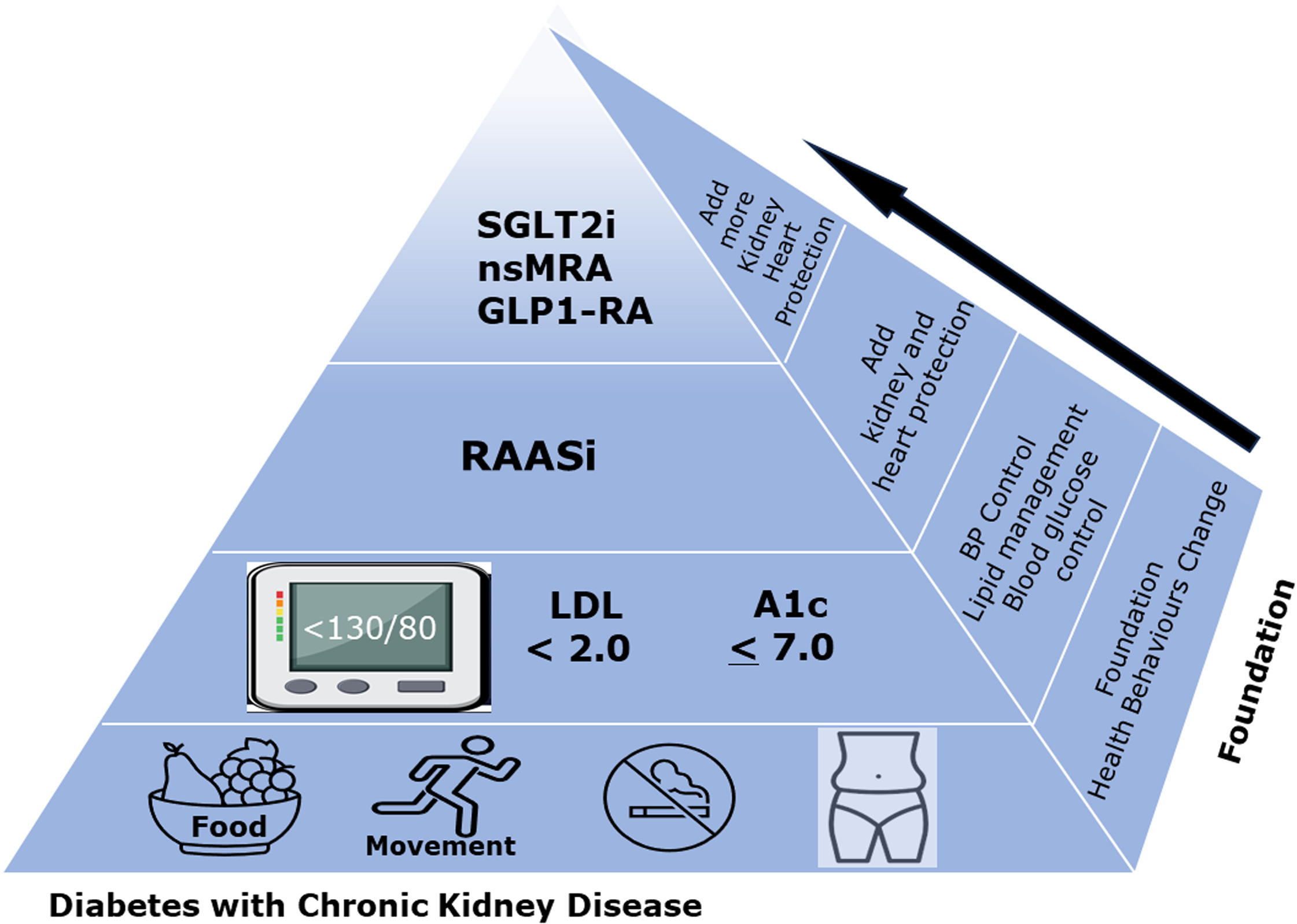
A graphical summary of an approach based on the recommendations from the working group adapted from the 2022 KDIGO clinical practice guideline for the management of CKD is shown in Figure 6 [81]. Starting with a foundation of health behaviours designed to prevent or slow the onset of diabetes and continuing after the development of diabetes will help with medical therapy to delay target-organ damage. Initial priorities would include BP management, starting with a RAASi, blood glucose management, and lipid management. If urine albumin excretion remains abnormal, further therapy (based on individual preferences) with an SGLT2i, nsMRA, and/or GLP1-RA with proven kidney protection will help slow CKD progression and reduce cardiovascular outcomes (Figure 6).
Figure 6
Management of individuals with diabetic kidney disease with an emphasis on blood pressure, blood glucose, and dyslipidemia management, in addition to RAASis and newer recommended therapies. RAASi, renin-angiotensin-aldosterone system inhibitor. Adapted from KDIGO 2022 [81].
Management of Hyperkalemia in People With Diabetes and CKD
People with diabetes and CKD are at high risk for hyperkalemia [82,83]. Initiation and titration of RAAS blockers can also lead to hyperkalemia. The incidence of hyperkalemia has ranged from 3% to 7% in clinical trials with RAAS blockage monotherapy and was higher overall in trials with MRAs [87]. An episode of hyperkalemia increases the risk for a second episode [84]. In individuals with CKD and/or chronic heart failure receiving RAAS blockers, predictors of recurrent hyperkalemia within 6 months of the first episode include initial hyperkalemia with a potassium (K) level of ≥5.6 mmol/L, an eGFR of <60 mL/min per 1.73 m², diabetes, and use of spironolactone [85].
Hyperkalemia is associated with an increased risk for mortality, cardiovascular events, hospitalizations, and intensive care unit admissions [86]. Therefore, potassium (K) level should be checked between 1 and 2 weeks after initiation or titration of RAASis [87] and after 4 weeks for nsMRAs [17] in people with diabetes and CKD. In people with a rise in potassium (K) level of >30%, follow-up testing should be performed. Because of the cardiorenal benefits of RAASis, if these medications must be temporarily reduced or withheld due to hyperkalemia, a retrial of RAASi should be considered, potentially at a lower dose once hyperkalemia is resolved.
Individuals on RAASi treatment should receive education on appropriate dietary K intake and supplements, medications that affect K levels, and the importance of adhering to scheduled blood tests after initiation or titration of these medications. Mild hyperkalemia can be managed with dietary counselling by eliminating foods that contribute to acute hyperkalemia, such as bananas, oranges or orange juice, tomatoes or tomato juice, and highly refined foods. For moderate hyperkalemia, urinary K excretion should be increased by administration of diuretics, such as furosemide, and sodium bicarbonate in individuals with metabolic acidosis. In addition, increasing elimination of K through the gastrointestinal tract can be achieved with K binders. Sodium polystyrene sulfonate is an older binder with limited supportive evidence [88]. This medication has been associated with risk of acute bowel necrosis, hypernatremia, diarrhea, and gastrointestinal toxicity [89]. The newer binders, sodium zirconium cyclosilicate, and patiromer, facilitate optimization of RAAS blockage therapy and have supportive evidence. These medications have demonstrated safety and are generally better tolerated [84]. For hyperkalemia, despite the measures just noted, referral to a nephrologist is suggested. For severe hyperkalemia, it is recommended to hold RAAS blockers and MRAs [90], and an emergency room referral or an emergency medical service call, depending on the individual clinical situation, is suggested (Table 8). Further information is presented in the supplementary material.
| Table 8 Classification of hyperkalemia |
|
|---|---|
| Severity of hyperkalemia | Serum K level (mmol/L) |
| Mild | Over the upper limit of normal: 5.4 |
| Moderate | 5.5–5.9 |
| Severe | ≥6.0 |
| K, potassium. | |
Values and Preferences
The kidney and cardiovascular benefits of additional therapy with RAASi, SGLT2i, GLP1-RA, and nsMRA, including additional testing and visits, must be balanced against the need for additional medications, as well as associated lab testing for individuals with residual risk due to persistent nephropathy with A3 albuminuria. If possible, decision support tools should be used to help individuals understand the risk and benefits and assist with decision-making. We recognize that financial considerations will limit access to therapy and encourage health-care providers to advocate for the individuals in their care until these agents are funded. We believe that all people at risk of kidney disease from diabetes should be made aware of the latest therapies to allow for shared decision-making.
Multiple drug therapy and the concept of goal-directed medical therapy, as in the management of heart failure, should now be considered for management of people with diabetes and persistent nephropathy. Combining drug therapies has been shown to be of benefit. The use of risk prediction equations in clinical practice can enhance shared decision-making about the use of multiple therapies, and allow providers and individuals to balance risk, cost, and side effects, rather than relying on summary effects from clinical trials and meta-analyses.
Recommendations
- Individuals with diabetes and no history of kidney disease should be screened annually with both eGFR and random urine ACR to identify individuals with CKD. For individuals with type 1 diabetes, CKD screening should begin 5 years after onset or, if onset is at an early age, screening should start after puberty. For type 2 diabetes, CKD screening should begin at diagnosis and annually thereafter (see screening algorithm in Figure 1) [Grade D, Consensus].
- For individuals with diabetes, the ACR threshold for diagnosing A2 albuminuria is 2.0 mg/mmol [30,31,34] [Grade C, Level 3].
- Individuals with diabetes and CKD should have their eGFR and spot uACR assessed at least annually with more frequent testing (every 3 to 6 months) when eGFR is <60 mL/min per 1.73 m² or if uACR is >20 mg/mmol [22–24,58,75,76] [Grade 1A, Level 2].
- For individuals with diabetes, a combined creatinine/cystatin C estimate of eGFR should be considered in settings where the creatinine-based eGFR may be unreliable for an individual (see Table 6) [21] [Grade D, Consensus].
- The KFRE is suggested for individuals with diabetes and CKD stages G3 to G5 for predicting the risk of end-stage kidney disease over 5 years [17,42,43] [Grade C, Level 3].
- Individuals with diabetes should be treated to meet a target systolic BP of <130 mmHg and a diastolic BP of <80 mmHg. These target BP levels are the same as the BP treatment thresholds [46] [Grade A, Level 1].
- Adults with diabetes and CKD with either hypertension or albuminuria should be recommended a RAASi (an ACEi or ARB) to delay progression of CKD [Grade A, Level 1A for ACEi use in type 1 and type 2 diabetes, and for ARB use in type 2 diabetes [91–95]; Grade D, Consensus for ARB use in type 1 diabetes] [15].
- Adults with type 2 diabetes and CKD defined by eGFR between 20 and 45 mL/min per 1.73 m² regardless of uACR, or eGFR between 45 and 90 mL/min per 1.73 m² with uACR >20 mg/mmol, on maximally tolerated, or maximally prescribed doses of RAASis, should be recommended an SGLT2i to delay progression of CKD and progression to dialysis, and to reduce likelihood of cardiovascular events [57,58,75,76] [Grade A, Level 1A].
- Adults with type 2 diabetic nephropathy defined by eGFR between 25 and 50 mL/min per 1.73 m² with uACR between 10 and 500 mg/mmol, or if eGFR is >50 mL/min per 1.73 m² with uACR between 30 and 500 mg/mmol, on maximally tolerated, or maximally prescribed doses of RAASi, should be recommended a GLP1-RA with proven kidney benefit to reduce proteinuria and risk of worsening kidney function [72] [Grade A, Level 1A for subcutaneous semaglutide].
- Adults with type 2 diabetic nephropathy defined by eGFR between 25 and 90 mL/min per 1.73 m² with uACR between 3 and 30 mg/mmol with or without diabetic retinopathy, for those with eGFR between 25 and 60 mL/min per 1.73 m², or if eGFR is >25 mL/min per 1.73 m² with uACR between 30 and 500 mg/mmol, on maximally tolerated, or maximally prescribed doses of RAASi, with serum potassium ≤4.8 mmol/L, should be recommended a nsMRA with proven efficacy alongside potassium monitoring to improve kidney and cardiovascular outcomes [64,66,70,71] [Grade A, Level 1A for finerenone].
- For adults with diabetes, mild hyperkalemia (defined by potassium levels between the upper limit of normal and 5.4 mmol/L) can be managed with dietary intervention. For moderate hyperkalemia (defined by K levels between 5.4 and 5.9 mmol/L), it is recommended to initiate medical therapy to increase K excretion through the gastrointestinal tract or in the urine. For severe hyperkalemia (defined by potassium ≥6.0 mmol/L), it is suggested to hold RAASi and nsMRA medications and refer to an emergency room for management [Grade D, Consensus].
Acknowledgments
The authors thank the external reviewers, Allan Grill MD, CCFP (COE), MPH, FCFP, CCPE and Kelly Picard PhD, RD, CDE, for their insightful feedback and for the lending of their time and expertise.
Author Disclosures
S.T. is a participant in the Living Kidney Donor Safety Study sponsored by CIHR, a volunteer board member of the American Hypertension Specialists Certification Program, co-chair of C-CHANGE, Physician Organization Chair for CHEP+, and reports honoraria and speaker fees from AstraZeneca, Bayer, Boehringer Ingelheim, CHEP+, Eisai, GSK, Janssen, KMH, Novo Nordisk, and Otsuka. P.M. reports consulting fees from Alexion, Amgen, AstraZeneca, Bayer, Boehringer Ingelheim, GSK, Janssen, Lilly, Novartis, and Otsuka; grants and research funding from Alnylam, AstraZeneca, Bayer, Boehringer Ingelheim, GSK, Janssen, Medtronic, Novartis, and Otsuka; and speaker fees for Alexion, Astra-Zeneca, Bayer, Boehringer Ingelheim, GSK, Janssen, Medtronic, Novartis, and Otsuka. H.S.B. reports trial fees paid to his institution by Amgen, AstraZeneca, Anji, Biomea, Boehringer Ingelheim, Eli Lilly, GlaxoSmithKline, Ionis, Kowa, Novartis, Novo Nordisk, and Pfizer. N.T. reports grants from AstraZeneca, Bayer, Boehringer Ingelheim, Eli Lilly and Company, and Otsuka Pharmaceutical Co. Ltd.; consulting fees from Bayer, AstraZeneca, Boehringer Ingelheim, GSK, Otsuka Pharmaceutical Co. Ltd., ProKidney, Roche, Vera, and Eli Lilly and Company; and speaker fees from AstraZeneca, Bayer, Boehringer Ingelheim, GSK, Otsuka Pharmaceutical Co. Ltd., ProKidney, Roche, Vera, GSK, and Eli Lilly and Company. R.J. and T.P. have no conflicts to disclose.
References
- Caramori ML, Rossing P. Diabetic Kidney Disease. [Updated 2022 Aug 3]. In: Feingold KR, Anawalt B, Blackman MR, et al., editors. Endotext [Internet]. South Dartmouth (MA): MDText.com, Inc.; 2000.
- Eknoyan G. A historical overview of diabetic nephropathy. In: Roelofs JJ, Vogt L, editors. Diabetic Nephropathy: Pathophysiology and Clinical Aspects. Berlin: Springer; 2018.
- Watkins P, Blainey J, Brewer D, Fitzgerald M, Malins J, O’Sullivan D, et al. The natural history of diabetic renal disease: A follow-up study of a series of renal biopsies. Q J Med 1972;41:437e56.
- Scilletta S, Di Marco M, Miano N, Filippello A, Di Mauro S, Scamporrino A, et al. Update on diabetic kidney disease (DKD): Focus on non-albuminuric DKD and cardiovascular risk. Biomolecules 2023;13:752.
- Ekinci EI, Jerums G, Skene A, Crammer P, Power D, Cheong KY, et al. Renal structure in normoalbuminuric and albuminuric patients with type 2 diabetes and impaired renal function. Diabetes Care 2013;36:3620e6.
- Coresh J, Selvin E, Stevens LA, Manzi J, Kusek JW, Eggers P, et al. Prevalence of chronic kidney disease in the United States. JAMA 2007;298:2038e47.
- Hill NR, Fatoba ST, Oke JL, Hirst JA, O’Callaghan CA, Lasserson DS, et al. Global prevalence of chronic kidney disease—a systematic review and meta-analysis. PLoS One 2016;11:e0158765.
- Patel A, MacMahon S, Chalmers J, Neal B, Woodward M, Billot L, et al. Effects of a fixed combination of perindopril and indapamide on macrovascular and microvascular outcomes in patients with type 2 diabetes mellitus (the ADVANCE trial): A randomised controlled trial. Lancet 2007;370:829e40.
- Holman RR, Paul SK, Bethel MA, Matthews DR, Neil HA. 10-year follow-up of intensive glucose control in type 2 diabetes. N Engl J Med 2008;359:1577e89.
- Agarwal R. Pathogenesis of diabetic nephropathy. In: Chronic Kidney Disease and Type 2 Diabetes. Arlington (VA): American Diabetes Association, 2021.
- Tarnow L, Rossing P, Gall M-A, Nielsen FS, Parving H-H. Prevalence of arterial hypertension in diabetic patients before and after the JNC-V. Diabetes Care 1994;17:1247e51.
- Epstein M, Sowers JR. Diabetes mellitus and hypertension. Hypertension 1992;19:403e18.
- Cheung AK, Chang TI, Cushman WC, Furth SL, Hou FF, Ix JH, et al. Executive summary of the KDIGO 2021 clinical practice guideline for the management of blood pressure in chronic kidney disease. Kidney Int 2021;99:559e69.
- Gaede P, Vedel P, Larsen N, Jensen GV, Parving HH, Pedersen O. Multifactorial intervention and cardiovascular disease in patients with type 2 diabetes. N Engl J Med 2003;348:383e93.
- Diabetes Canada Clinical Practice Guidelines Expert Committee. Chronic kidney disease in diabetes. Can J Diabetes 2018;42(Suppl):S201e9.
- Lipscombe L, Butalia S, Dasgupta K, Eurich DT, MacCallum L, Shah BR, et al. Pharmacologic glycemic management of type 2 diabetes in adults: 2020 update. Can J Diabetes 2020;44:575e91.
- Stevens PE, Ahmed SB, Carrero JJ, Foster B, Francis A, Hall RK, et al. KDIGO 2024 clinical practice guideline for the evaluation and management of chronic kidney disease. Kidney Int 2024;105(Suppl.):S117e314.
- Kidney Disease: Improving Global Outcomes Blood Pressure Work Group. KDIGO 2021 clinical practice guideline for the management of blood pressure in chronic kidney disease. Kidney Int 2021;99(Suppl.):S1e87.
- Go AS, Chertow GM, Fan D, McCulloch CE, Hsu CY. Chronic kidney disease and the risks of death, cardiovascular events, and hospitalization. N Engl J Med 2004;351:1296e305.
- Tobe SW, Clase CM, Gao P, McQueen M, Grosshennig A, Wang X, et al. Cardiovascular and renal outcomes with telmisartan, ramipril, or both in people at high renal risk: Results from the ONTARGET and TRANSCEND studies. Circulation 2011;123(10):1098-107.
- Inker LA, Titan S. Measurement and estimation of GFR for use in clinical practice: Core curriculum 2021. Am J Kidney Dis 2021;78:736e49.
- Lindeman RD, Tobin J, Shock NW. Longitudinal studies on the rate of decline in renal function with age. J Am Geriatr Soc 1985;33:278e85.
- Yu Z, Grams ME, Ndumele CE, Wagenknecht L, Boerwinkle E, North KE, et al. Association between midlife obesity and kidney function trajectories: The Atherosclerosis Risk in Communities (ARIC) study. Am J Kidney Dis 2021;77: 376e85.
- Madero M, Katz R, Murphy R, Newman A, Patel K, Ix J, et al. Comparison between different measures of body fat with kidney function decline and incident CKD. Clin J Am Soc Nephrol 2017;12:893e903.
- Herold JM, Wiegrebe S, Nano J, Jung B, Gorski M, Thorand B, et al. Population- based reference values for kidney function and kidney function decline in 25- to 95-year-old Germans without and with diabetes. Kidney Int 2024;106: 699e711.
- Kouri TT, Viikari JS, Mattila KS, Irjala KM. Microalbuminuria. Invalidity of simple concentration-based screening tests for early nephropathy due to urinary volumes of diabetic patients. Diabetes Care 1991;14:591e3.
- Ahn CW, Song YD, Kim JH, Lim SK, Choi KH, Kim KR, et al. The validity of random urine specimen albumin measurement as a screening test for diabetic nephropathy. Yonsei Med J 1999;40:40e5.
- Bakker AJ. Detection of microalbuminuria. Receiver operating characteristic curve analysis favors albumin-to-creatinine ratio over albumin concentration. Diabetes Care 1999;22:307e13.
- Clinical Practice Guidelines Expert Committee. Canadian Diabetes Association 2003 clinical practice guidelines for the prevention and management of diabetes in Canada. Nephropathy. Can J Diabetes 2003;27(Suppl. 2): S66e71.
- Matsushita K. Association of estimated glomerular filtration rate and albuminuria with all-cause and cardiovascular mortality in general population cohorts: A collaborative meta-analysis. Lancet 2010;375:2073e81.
- Fox CS, Matsushita K, Woodward M, Bilo HJG, Chalmers J, Heerspink HJL, et al. Associations of kidney disease measures with mortality and end-stage renal disease in individuals with and without diabetes: A meta-analysis. Lancet 2012;380:1662e73.
- Keane WF, Brenner BM, de Zeeuw D, Grunfeld JP, McGill J, Mitch WE, et al. The risk of developing end-stage renal disease in patients with type 2 diabetes and nephropathy: The RENAAL study. Kidney Int 2003;63:1499e507.
- Mann JF, Gerstein HC, Yi QL, Lonn EM, Hoogwerf BJ, Rashkow A, et al. Development of renal disease in people at high cardiovascular risk: Results of the HOPE randomized study. J Am Soc Nephrol 2003;14:641e7.
- Ruggenenti P, Porrini E, Motterlini N, Perna A, Ilieva AP, Iliev IP, et al. Measurable urinary albumin predicts cardiovascular risk among normoalbuminuric patients with type 2 diabetes. J Am Soc Nephrol 2012;23:1717e24.
- Smekal MD, Tam-Tham H, Finlay J, Donald M, Thomas C, Weaver RG, et al. Patient and provider experience and perspectives of a risk-based approach to multidisciplinary chronic kidney disease care: A mixed methods study. BMC Nephrol 2019;20:110.
- Che M, Iliescu E, Thanabalasingam S, Day AG, White CA. Death and dialysis following discharge from chronic kidney disease clinic: A retrospective cohort study. Can J Kidney Health Dis 2022;9:20543581221118434.
- Grams ME, Brunskill NJ, Ballew SH, Sang Y, Coresh J, Matsushita K, et al. The kidney failure risk equation: Evaluation of novel input variables including eGFR estimated using the CKD-EPI 2021 equation in 59 cohorts. J Am Soc Nephrol 2023;34:482e94.
- Kang MW, Tangri N, Kim YC, An JN, Lee J, Li L, et al. An independent validation of the kidney failure risk equation in an Asian population. Sci Rep 2020;10: 12920.
- Lennartz CS, Pickering JW, Seiler-Mussler S, Bauer L, Untersteller K, Emrich IE, et al. External validation of the kidney failure risk equation and re-calibration with addition of ultrasound parameters. Clin J Am Soc Nephrol 2016;11: 609e15.
- Ooi Y-G, Sarvanandan T, Hee NKY, Lim Q-H, Paramasivam SS, Ratnasingam J, et al. Risk prediction and management of chronic kidney disease in people living with type 2 diabetes mellitus. Diabetes Metab J 2024;48(2):196e207.
- Tangri N, Ferguson T, Leon SJ, Anker SD, Filippatos G, Pitt B, et al. Validation of the Klinrisk chronic kidney disease progression model in the FIDELITY population. Clin Kidney J 2024;17:sfae052.
- Tangri N, Ferguson TW, Wiebe C, Eng F, Nash M, Astor BC, et al. Validation of the kidney failure risk equation in kidney transplant recipients. Can J Kidney Health Dis 2020;7:2054358120922627.
- Whitlock RH, Chartier M, Komenda P, Hingwala J, Rigatto C, Walld R, et al. Validation of the kidney failure risk equation in Manitoba. Can J Kidney Health Dis 2017;4:2054358117705372.
- National Institute for Health and Care Excellence. Evidence Review for the Best Combination of Measures to Identify Increased Risk of Progression in Adults, Children and Young People: Chronic Kidney Disease: Evidence Review. London: NICE, 2021.
- Tangri N. Risk stratification to improve care and outcomes in diabetic kidney disease. Can Diabetes Endocrinol Today 2024;2:5e10.
- Can Diabetes Endocrinol Today 2024;2:5e10. Rabi DM, McBrien KA, Sapir-Pichhadze R, Nakhla M, Ahmed SB, Dumanski SM, et al. Hypertension Canada’s 2020 comprehensive guidelines for the prevention, diagnosis, risk assessment, and treatment of hypertension in adults and children. Can J Cardiol 2020;36:596e624.
- Mogensen C. Long-term antihypertensive treatment inhibiting progression of diabetic nephropathy. BMJ (Clin Res Ed) 1982;285:685e8.
- Dagenais GR, Yusuf S, Bourassa MG, Yi Q, Bosch J, Lonn EM, et al. Effects of ramipril on coronary events in high-risk persons. Circulation 2001;104:522e6.
- Tobe SW, Clase CM, Gao P, McQueen M, Grosshennig A, Wang X, et al. Cardiovascular and renal outcomes with telmisartan, ramipril, or both in people at high renal risk: Results from the ONTARGET and TRANSCEND studies. Circulation 2011;123:1098e107.
- Abdul-Ghani MA, Norton L, DeFronzo RA. Renal sodium-glucose cotransporter inhibition in the management of type 2 diabetes mellitus. Am J Physiol 2015; 309:F889e900.
- Cherney DZI, Cooper ME, Tikkanen I, Pfarr E, Johansen OE, Woerle HJ, et al. Pooled analysis of Phase III trials indicate contrasting influences of renal function on blood pressure, body weight, and HbA1c reductions with empagliflozin. Kidney Int 2018;93:231e44.
- Zinman B, Wanner C, Lachin JM, Fitchett D, Bluhmki E, Hantel S, et al. Empagliflozin, cardiovascular outcomes, and mortality in type 2 diabetes. N Engl J Med 2015;373:2117e28.
- Neal B, Perkovic V, Mahaffey KW, de Zeeuw D, Fulcher G, Erondu N, et al. Canagliflozin and cardiovascular and renal events in type 2 diabetes. N Engl J Med 2017;377:644e57.
- Wanner C, Inzucchi SE, Lachin JM, Fitchett D, von Eynatten M, Mattheus M, et al. Empagliflozin and progression of kidney disease in type 2 diabetes. N Engl J Med 2016;375:323e34.
- Perkovic V, Jardine MJ, Neal B, Bompoint S, Heerspink HJL, Charytan DM, et al. Canagliflozin and renal outcomes in type 2 diabetes and nephropathy. N Engl J Med 2019;380:2295e306.
- Heerspink HJL, Stefansson BV, Correa-Rotter R, Chertow GM, Greene T, Hou FF, et al. Dapagliflozin in patients with chronic kidney disease. N Engl J Med 2020; 383:1436e46.
- The E-KCG, Herrington WG, Staplin N, Wanner C, Green JB, Hauske SJ, et al. Empagliflozin in patients with chronic kidney disease. N Engl J Med 2023;388: 117e27.
- Herrington W, Staplin N, Haynes R, Mayne K, Roddick A, Landray M, et al. Impact of diabetes on the effects of sodium glucose co-transporter-2 (SGLT2) inhibitors on kidney outcomes: Collaborative meta-analysis of large placebocontrolled trials. Lancet 2022;400.
- Lewis EJ, Hunsicker LG, Clarke WR, Berl T, Pohl MA, Lewis JB, et al. Renoprotective effect of the angiotensin-receptor antagonist irbesartan in patients with nephropathy due to type 2 diabetes. N Engl J Med 2001;345:851e60.
- Brenner BM, Cooper ME, de Zeeuw D, Keane WF, Mitch WE, Parving HH, et al. Effects of losartan on renal and cardiovascular outcomes in patients with type 2 diabetes and nephropathy. N Engl J Med 2001;345:861e9.
- McGovern AP, Hogg M, Shields BM, Sattar NA, Holman RR, Pearson ER, et al. Risk factors for genital infections in people initiating SGLT2 inhibitors and their impact on discontinuation. BMJ Open Diabetes Res Care 2020;8: e001238.
- Colacci M, Fralick J, Odutayo A, Fralick M. Sodium-glucose cotransporter-2 inhibitors and risk of diabetic ketoacidosis among adults with type 2 diabetes: A systematic review and meta-analysis. Can J Diabetes 2022;46:10e15.e2.
- Tobe SW, Mavrakanas TA, Bajaj HS, Levin A, Tangri N, Slee A, et al. Impact of canagliflozin on kidney and cardiovascular outcomes by type 2 diabetes duration: A pooled analysis of the CANVAS program and CREDENCE trials. Diabetes Care; 2024:dc231450.
- van Raalte DH, Bjornstad P, Cherney DZ, de Boer IH, Fioretto P, Gordin D, et al. Combination therapy for kidney disease in people with diabetes mellitus. Nat Rev Nephrol; 2024:1e14.
- Wu Y, Lin H, Tao Y, Xu Y, Chen J, Jia Y, et al. Network meta-analysis of mineralocorticoid receptor antagonists for diabetic kidney disease. Front Pharmacol 2022;13:967317.
- Dutta D, Surana V, Bhattacharya S, Aggarwal S, Sharma M. Efficacy and safety of novel non-steroidal mineralocorticoid receptor antagonist finerenone in the management of diabetic kidney disease: A meta-analysis. Ind J Endocrinol Metabol 2022;26:198e205.
- Pitt B, Filippatos G, Agarwal R, Anker SD, Bakris GL, Rossing P, et al. Cardiovascular events with finerenone in kidney disease and type 2 diabetes. N Engl J Med 2021;385:2252e63.
- Bakris GL, Agarwal R, Anker SD, Pitt B, Ruilope LM, Rossing P, et al. Effect of finerenone on chronic kidney disease outcomes in type 2 diabetes. N Engl J Med 2020;383:2219e29.
- Ruilope LM, Agarwal R, Anker SD, Bakris GL, Filippatos G, Nowack C, et al. Design and baseline characteristics of the finerenone in reducing cardiovascular mortality and morbidity in diabetic kidney disease trial. Am J Nephrol 2019;50:345e56.
- Agarwal R, Filippatos G, Pitt B, Anker SD, Rossing P, Joseph A, et al. Cardiovascular and kidney outcomes with finerenone in patients with type 2 diabetes and chronic kidney disease: The FIDELITY pooled analysis. Eur Heart J 2022;43:474e84.
- Zheng Y, Ma S, Huang Q, Fang Y, Tan H, Chen Y, et al. Meta-analysis of the efficacy and safety of finerenone in diabetic kidney disease. Kidney Blood Pressure Res 2022;47:219e28.
- Perkovic V, Tuttle KR, Rossing P, Mahaffey KW, Mann JF, Bakris G, et al. Effects of semaglutide on chronic kidney disease in patients with type 2 diabetes. N Engl J Med; 2024.
- Mann JF, Rossing P, Bakris G, Belmar N, Bosch-Traberg H, Busch R, et al. Effects of semaglutide with and without concomitant SGLT2 inhibitor use in participants withtype2 diabetesand chronic kidneydisease in the FLOWtrial. NatMed2024;1.
- Evans M, Bain SC, Hogan S, Bilous RW. Irbesartan delays progression of nephropathy as measured by estimated glomerular filtration rate: Post hoc analysis of the Irbesartan Diabetic Nephropathy Trial. Nephrol Dial Transplant 2011;27:2255e63.
- Perkovic V, Jardine MJ, Neal B, Bompoint S, Heerspink HJL, Charytan DM, et al. Canagliflozin and renal outcomes in type 2 diabetes and nephropathy. N Engl J Med 2019;380:2295e306.
- Heerspink HJ, Jongs N, Chertow GM, Langkilde AM, McMurray JJ, Correa- Rotter R, et al. Effect of dapagliflozin on the rate of decline in kidney function in patients with chronic kidney disease with and without type 2 diabetes: A prespecified analysis from the DAPA-CKD trial. Lancet Diabetes Endocrinol 2021;9:743e54.
- Bakris GL, Ruilope LM, Anker SD, Filippatos G, Pitt B, Rossing P, et al. A prespecified exploratory analysis from FIDELITY examined finerenone use and kidney outcomes in patients with chronic kidney disease and type 2 diabetes. Kidney Int 2023;103:196e206.
- Yang S, Zhao L, Mi Y, He W. Effects of sodium-glucose cotransporter-2 inhibitors and aldosterone antagonists, in addition to renin-angiotensin system antagonists, on major adverse kidney outcomes in patients with type 2 diabetes and chronic kidney disease: A systematic review and network meta-analysis. Diabetes Obes Metabol 2022;24:2159e68.
- Ghosal S, Sinha B. Assessing the effects of modern renoprotective agents in preventing progression of renal composite outcomes in patients with type 2 diabetes: A network meta-analysis. Diabetes Ther 2023;14:415e24.
- Neuen BL, Heerspink HJ, Vart P, Claggett BL, Fletcher RA, Arnott C, et al. Estimated lifetime cardiovascular, kidney, and mortality benefits of combination treatment with SGLT2 inhibitors, GLP-1 receptor agonists, and nonsteroidal MRA compared with conventional care in patients with type 2 diabetes and albuminuria. Circulation 2024;149:450e62.
- KDIGO. KDIGO 2022 clinical practice guideline for diabetes management in chronic kidney disease. Kidney Int 2022;102(Suppl.):S1e127.
- Desai AS, Swedberg K, McMurray JJ, Granger CB, Yusuf S, Young JB, et al. Incidence and predictors of hyperkalemia in patients with heart failure: An analysis of the CHARM program. J Am Coll Cardiol 2007;50:1959e66.
- Pitt B, Bakris G, Ruilope LM, DiCarlo L, Mukherjee R. Serum potassium and clinical outcomes in the Eplerenone Post-Acute Myocardial Infarction Heart Failure Efficacy and Survival Study (EPHESUS). Circulation 2008;118: 1643e50.
- Weinstein J, Girard L-P, Lepage S, McKelvie RS, Tennankore K. Prevention and management of hyperkalemia in patients treated with renineangiotensinealdosterone system inhibitors. CMAJ 2021;193: E1836e41.
- Adelborg K, Nicolaisen SK, Hasvold P, Palaka E, Pedersen L, Thomsen RW. Predictors for repeated hyperkalemia and potassium trajectories in high risk patients—a population-based cohort study. PLoS One 2019;14: e0218739.
- Hougen I, Leon SJ, Whitlock R, Rigatto C, Komenda P, Bohm C, et al. Hyperkalemia and its association with mortality, cardiovascular events, hospitalizations, and intensive care unit admissions in a population-based retrospective cohort. Kidney Int Rep 2021;6:1309e16.
- Miyamori I, Yasuhara S, Takeda Y, Koshida H, Ikeda M, Nagai K, et al. Effects of converting enzyme inhibition on split renal function in renovascular hyper-tension. Hypertension 1986;8:415e21.
- Lepage L, Dufour A-C, Doiron J, Handfield K, Desforges K, Bell R, et al. Randomized clinical trial of sodium polystyrene sulfonate for the treatment of mild hyperkalemia in CKD. Clin J Am Soc Nephrol 2015;10:2136e42.
- Pitt B, Bakris GL. New potassium binders for the treatment of hyperkalemia: Current data and opportunities for the future. Hypertension 2015;66:731e8.
- Palmer BF. Managing hyperkalemia caused by inhibitors of the renineangiotensinealdosterone system. N Engl J Med 2004;351:585e92.
- Lewis EJ, Hunsicker LG, Bain RP, Rohde RD. The effect of angiotensin- converting-enzyme inhibition on diabetic nephropathy. N Engl J Med 1993; 329:1456e62.
- Lewis EJ, Hunsicker LG, Clarke WR, Berl T, Pohl MA, Lewis JB, et al. Reno-protective effect of the angiotensin-receptor antagonist irbesartan in patients with nephropathy due to type 2 diabetes. N Engl J Med 2001;345:851e60.
- Brenner BM, Cooper ME, de Zeeuw D, Keane WF, Mitch WE, Parving HH, et al. Effects of losartan on renal and cardiovascular outcomes in patients with type 2 diabetes and nephropathy. N Engl J Med 2001;345:861e9.
- Parving HH, Lehnert H, Brochner-Mortensen J, Gomis R, Andersen S, Arner P, et al. The effect of irbesartan on the development of diabetic nephropathy in patients with type 2 diabetes. N Engl J Med 2001;345:870e8.
- ACE Inhibitors in Diabetic Nephropathy Trialist Group. Should all patients with type 1 diabetes mellitus and microalbuminuria receive angiotensin-converting enzyme inhibitors? A meta-analysis of individual patient data. Ann Intern Med 2001;134:370e9.
Hyperkalemia
Introduction
In clinical practice, hyperkalemia is one of the most important electrolyte disorders [1]. This disorder is associated with an increased risk for mortality, cardiovascular events, hospitalizations, and intensive care unit admissions [2], and is more common in individuals with chronic kidney disease (CKD) and diabetes treated with guideline-directed medical therapy [3]. One of these therapies is renin–angiotensin–aldosterone system inhibition (RAASi), which has been shown to increase hyperkalemia risk [4].
Unfortunately, clinicians often react to an episode of hyperkalemia by reducing or discontinuing RAASi [5]. Studies have demonstrated that both discontinuation and submaximal doses of RAASi are associated with an elevated risk of cardiorenal morbidity and mortality [6]. Therefore, our goal is to continue RAASi as part of guideline-directed medical therapy and to protect individuals at risk from hyperkalemia. This supplementary section provides clinicians with practical tools, strategies, and recommendations for interprofessional management of hyperkalemia of adult outpatients with CKD and diabetes who have a compelling indication for RAASi therapy. In what follows, RAASi refers to angiotensin-converting enzyme inhibitors (ACEis), angiotensin receptor blockers (ARBs), and mineralocorticoid receptor antagonists (MRAs).
Definition of Hyperkalemia
There is no universally agreed-upon definition for hyperkalemia. The most common definition is a potassium (K) level exceeding 5.0 mmol/L. However, due to the variability of the upper limit of normal (ULN) for potassium levels reported by the different laboratories (labs) in Canada, we define hyperkalemia as a potassium level above the ULN as recorded in an individual’s lab report.
Hyperkalemia is a K level above the ULN as recorded in an individual’s lab report.
Note: Potassium levels are measured in serum or in plasma. In our review, we found that many studies and reviewed articles did not clearly state whether serum or plasma potassium was measured. For simplicity, we use the general expression “K level” in this document. The symbol 'K' for potassium comes from the Latin word Kalium.
Classification of Hyperkalemia
Potassium thresholds for mild, moderate, and severe hyperkalemia differ among guidelines and in studies. We suggest the following definitions for our classification:
Practice Pearl
Mild hyperkalemia: K>ULN to 5.4 mmol/L
Moderate hyperkalemia: K=5.5 to 5.9 mmol/L
Severe hyperkalemia: K≥6 mmol/L
Our classification aligns with the Canadian Cardiovascular Society (CCS) Heart Failure classification [7] and the European Society of Cardiology classification (ESC) [8]. This classification differs from Alfonzo’s NICE (National Institute for Health and Care Excellence) guideline from the UK, where severe only starts at K = 6.5 mmol/L [9]. Our consensus conference’s choice of severe hyperkalemia starting at 6 mmol/L in 2024 was in part due to the arrival of new agents for treating hyperkalemia that are safe and effective, in contrast to the more complex legacy treatments with many side effects and a paucity of data to support them.
Overview of Potassium Homeostasis
In normal human physiology, potassium plays a key role in maintaining the ion concentration between the intra- and extracellular spaces, and the membrane potential of the cells [10]. Adults are typically composed of 60% water, two thirds inside cells and one third in the extracellular space, giving our skin turgor and allowing nutrients to flow from our blood vessels into cells. Ninety-eight percent of potassium is present inside our cells, and only 2% in the extracellular fluid space [11]. The extracellular fraction is essential for keeping the body in homeostasis or balance [10]. The addition of small amounts of potassium to the extracellular fluid over a short time can greatly increase the potassium concentration in the blood, resulting in electrophysiologic disturbances [1].
Maintaining normal potassium levels (Supplementary Figure 1) is the responsibility of several mechanisms, with the kidneys being of primary importance and responsible for excreting the daily dietary potassium intake minus what is lost through the gut [12]. Normally functioning kidneys also have a remarkable capacity to increase potassium excretion in the setting of excess potassium intake [13]. The process is relatively slow, and takes hours, and is also affected by diurnal variation, with less excretion overnight when urine flows tend to be low to allow sleep, and higher during the day [13]. If our recommended daily potassium intake ended up in our extracellular fluid all at once, potassium levels would more than double causing our circulatory system to fail. To prevent this, we have evolved to slowly move potassium from our digestive system through the extracellular fluid and into our cells. After a meal, catecholamines (the “fight-or-flight” hormones) and insulin are released and together promote potassium entry into cells, particularly the muscles and liver [12]. Potassium is then slowly released from the cells into the bloodstream, filtered by the kidneys, and excreted over many hours [12]. This process requires normally functioning kidneys making sufficient urine to transport all the potassium that needs to be excreted. Our bodies are highly regulated to ensure just the right amount of potassium is excreted.
Potassium excretion is regulated in large part by the RAAS, with aldosterone playing a major role in the kidney’s ability to excrete more potassium. It is, therefore, not surprising that blood pressure (BP)–lowering drugs that inhibit the RAAS system will slow potassium excretion, increasing potassium levels. The gastrointestinal (GI) system accounts for about 10% of total potassium excretion [12]. In CKD, the kidney’s ability to filter is reduced, lowering the amount of potassium that can be excreted, and, when urine flow is further reduced due to volume depletion or dehydration, potassium excretion is further limited. Also, the ability of insulin to stimulate potassium uptake into cells is also reduced in CKD from diabetes [14]. Over time, when excretion from the kidneys becomes limited, GI excretion of potassium is significantly enhanced, particularly in individuals undergoing dialysis [15], becoming an important potassium homeostatic mechanism that can also lead to high potassium levels if constipation occurs. It also presents an opportunity to increase potassium excretion with potassium binders that pass right through the GI system [16].
Prevalence, Incidence, and Risk of Hyperkalemia
The prevalence of hyperkalemia is variable depending on the population studied and the threshold at which hyperkalemia is diagnosed. A systematic review and meta-analysis of the incidence and prevalence of hyperkalemia in 527 observational studies of different populations from 63 countries reported the pooled mean prevalence of hyperkalemia (K>5.0 mmol/L) for adult populations with nondialysis CKD was 14.6% and with diabetes was 8.4%, respectively [17]. Very high potassium levels cause potentially fatal cardiac dysrhythmias. In a retrospective cohort study from Manitoba, hyperkalemia was associated with an increased risk for mortality, cardiovascular events, hospitalizations, and intensive care unit admissions [2]. Hyperkalemia was significantly associated with a 15% increased risk for all-cause mortality, a 71% hospitalization increase, and a more than tripled risk of admission to the intensive care unit [2]. Observational studies also found that new onset of chronic mild-to-moderate hyperkalemia (K=5.0 to 6.0 mmol/L) was associated with an increased risk of end-stage kidney disease by 30% [18,19]. It is, therefore, important to be aware of hyperkalemia and how to manage the condition.
A matched case-control study by Bakris et al of individuals with stage 3 or 4 CKD compared individuals with recurrent hyperkalemia over 12 months vs those who remained normokalemic after 5 years [20]. Over the 5-year follow-up, there was an all-cause increased mortality risk of 1.29-fold vs the normokalemia cohort (45% vs 37%), a 1.40-fold increased risk for major adverse cardiac events (MACEs) (60% vs 45%), a risk of 1.72 (34% vs 19%) for RAASi discontinuation, and a 1.94-fold higher risk of hospitalization with arrhythmia compared with the matched normokalemia cohort [20].
The fear that hyperkalemia will cause cardiac arrhythmias often prompts clinicians to stop or reduce kidney-protective therapy, including ACEis, or ARBs or MRAs, and if these drugs are not restarted, then people miss out on their kidney- and heart-protective benefits. In the hyperkalemia algorithm presented in this work, these drugs are not stopped unless K≥6 mmol/L. Data from a large retrospective US clinical care cohort study of 1,772,900 people with CKD (either low estimated glomerular filtration rate [eGFR] or abnormal albuminuria) representative of the general population was able to provide rates of hyperkalemia per 100 person years for different levels of eGFR and albuminuria, as well as by certain comorbidities (Supplementary Table 1) [21].
The median age of this cohort was 75 years, 57.7% were female, and 34% had diabetes. By comorbidity, including CKD, incident rates for hyperkalemia per 100 person years was 5.43 for people with type 2 diabetes, 8.7 for heart failure, 4.03 for people taking an ACEi or ARB, and 7.66 taking a steroidal MRA [21]. By comorbidity, hyperkalemia rises from 1 per 100 person years for people with normal eGFR and normal albuminuria to 19 per 100 person years, or almost 1 in 5 people per year for those with both stage G4 (severely decreased kidney function) and severe nephropathy (abnormal urine albumin levels).
These data show that the people who need treatment that causes hyperkalemia the most are those most likely to get hyperkalemia. This reinforces the message that they should be screened regularly—annually for very stable individuals and every 3 or 6 months if they are at higher risk. If, due to hyperkalemia, ACEi, ARB, or MRA must be held, it should be restarted again when potassium levels have fallen sufficiently.
Risk Factors for Hyperkalemia and Strategies for Management
An extensive review of all factors contributing to hyperkalemia is beyond the scope of this work. A brief discussion of common causes of hyperkalemia related to CKD and diabetes and their mechanisms is presented in what follows.
1. Common medical conditions
Two mechanisms contributing to hyperkalemia are abnormal transcellular shift of potassium and decreased/impaired potassium excretion (Supplementary Table 2). Metabolic acidosis causes potassium to shift from the intracellular space to the extracellular space [14]. With insulin deficiency or resistance, individuals may have difficulty moving potassium from the extracellular compartment to the intracellular space [1].
Individuals with CKD and diabetes often have additional medical conditions that increase their risk for hyperkalemia. Comorbidities such as heart failure and cardiovascular disease, prevalent in individuals with CKD and diabetes, increase the risk for hyperkalemia, usually due to associated medications. Chronic hyperkalemia is typically caused by reduced kidney clearance of potassium with more severe CKD. It is also worsened by reduced bowel motility and constipation, reducing the excretion of potassium by the GI tract, which becomes a larger share of total body excretion with CKD [22]. In diabetes, endocrine factors also increase potassium levels. Insulin resistance in type 2 diabetes and insulin deficiency in type 1 diabetes slow the movement of extracellular potassium into cells. Also, after many years of diabetes, the ability to increase kidney potassium excretion with aldosterone is reduced due to damage of the nerves innervating the kidneys, reducing renin and thus aldosterone levels [22–24].
Hyperkalemia also occurs rapidly. After an acute kidney injury (AKI) event, when kidney function suddenly falls close to or below the level necessary to sustain life, acute hyperkalemia results, worsened by metabolic acidosis. With metabolic acidosis (a reduction of the serum bicarbonate level), protons (acid) enter the cells and, to maintain electrical neutrality, potassium shifts from within the cells into the extracellular space. Acute increases in potassium are also commonly caused by ingestion of potassium-rich fruits and vegetables, particularly during the summer months when grocery stores are full of local tomatoes and other produce. Less commonly, hyperkalemia results from a GI bleed, where the red cells in the gut are digested, releasing their potassium, which is then quickly absorbed into the bloodstream. Concurrent volume depletion from the bleeding impairs potassium excretion from the kidney.
Practice Pearl
For the management of hyperkalemia, use all possible interventions to lower potassium level before discontinuing or dose reducing RAAS inhibitors.
2. Medications
Individuals with CKD and diabetes are often treated with various medications that increase the risk for hyperkalemia (Supplementary Table 3). Among these medications, RAAS inhibitors (ACEis and ARBs) provide cardiorenal benefit, but they also commonly contribute to hyperkalemia. After an episode of hyperkalemia, discontinuation or dose reduction of RAAS inhibitors is common [5]. An analysis of a large US database showed that, in individuals on RAASi therapy who developed hyperkalemia, the dose was titrated down in 1 of 5 hyperkalemic events and was discontinued in another 1 of 5 hyperkalemic events [26]. A Canadian population-based cohort study by Leon et al showed that, in Ontario, stopping RAASi therapy increased the risks of both overall death and need for dialysis [5].
3. Herbal products containing potassium
To our knowledge, authoritative information on herbal products with potassium is sparse. Numerous products have been known to affect both kidney function and electrolytes. Products such as alfalfa (Medicago sativa), dandelion (Taraxacum officinale), horse tail (Equisetum arvense), and nettle (Urticadioica) contain significant amounts of potassium and have been known to induce hyperkalemia in individuals with underlying risk factors [27]. Herbal products with a digitalis-like effect, including milkweed, lily of the valley, Siberian ginseng, and Hawthorn berries, reduce Na-K-ATPase activity, leading to elevated extracellular potassium [28]. In addition to inducing hyperkalemia, there is a concern that many herbal products have not been evaluated in individuals with CKD and may cause serious adverse effects [30]. Herbal products and their pharmacologically active ingredients can potentially cause nephrotoxicity, especially during volume depletion and acute illnesses and harmful drug interactions [31]. One review on plants and herbal components with antiarrhythmic activities and their interaction with cardiac medications reported multiple interactions, including effects on drugs known to impact potassium levels (Supplementary Figure 2), potentially enhancing the pro-arrhythmogenic effect of cardiovascular medications [32].
- Review medications/over-the-counter products with individuals regularly.
- Advise individuals to avoid herbal products to prevent risk of hyperkalemia and drug interactions.
- Monitor kidney function and volume status regularly in individuals who are using herbal products.
The National Kidney Foundation (NKF) recommends caution for a number of herbal supplements known to include potassium (National Kidney Foundation, https://www.kidney.org/atoz/content/herbalsupp#which-herbal-supplements-have-potassium, accessed November 11, 2025) (Supplementary Table 4).
4. Nutrition
Dietary potassium is mostly absorbed in the duodenum and jejunum, with a net intestinal absorption of approximately 90% [22]. There are 3 main factors contributing to daily dietary potassium intake: dietary potassium, potassium-enriched salt substitutes, and potassium additives. The following subsections address their management.
(a) Diet
Dietary potassium restriction in individuals with CKD has been a core strategy since the 1960s [23,33]. However, this strategy is supported by limited data [24,34]. Study data for the safety of increased potassium intake or liberalization of potassium restrictions in individuals with advanced CKD is also lacking. Nephrology programs are good resources for information about dietary potassium intake with hyperkalemia [24].
For the early stages of CKD, a recent systematic review of dietary potassium intake and risk of CKD progression suggested that higher dietary potassium intake was protective against disease progression, and that dietary restriction of foods naturally containing potassium may be harmful to cardiac health [35]. Although well-designed clinical trials are required to ascertain the risk/benefit of plant-based diets, emerging evidence has questioned the practices of severely limiting potassium intake from fruits and vegetables over the years to reduce the risk of and treatment of hyperkalemia [23]. Furthermore, dietary potassium from animal sources (dairy, meats, and soups), highly processed foods, and foods with potassium additives are more likely to have greater impact on potassium level than the potassium from plant-based foods. This is due to greater bioavailability and faster potassium absorption rates from highly processed foods (Supplementary Figure 3) [36]. The 2024 KDIGO guidelines recommend limiting the intake of foods rich in bioavailable potassium for individuals with stage 3 to 5 CKD who have a history of hyperkalemia or as a preventive strategy during periods in which hyperkalemia risk may be a concern [30].
The diets prescribed for people with CKD, diabetes, and hyperkalemia are quite complex and challenging to adhere to as these individuals often have additional dietary restrictions from concurrent comorbidities (e.g. low-sodium diet and reduced fluid intake for heart failure, or carbohydrate limits for diabetes, fat limits for heart disease, and protein limits for kidney disease). Healthy nutritional plans should include factors such as potassium and other nutrient bioavailability, cooking methods, portion sizes, food combinations, fibre content, dietary acid load, and comorbidities [34], and should reflect each individual’s cultural values/beliefs and preferences for better adherence. Dietitians are therefore well-positioned and necessary to provide individualized dietary recommendations.
Practice Pearl
For prevention of or management of hyperkalemia, refer to the renal dietitian.
Practice Pearl
When a diet consultation with a registered dietitian is not immediately available, following Supplementary Figures 4 and 5 and Supplementary Table 5 may be helpful for a quick review of nutrition/education with the individual.
(b) Potassium-enriched salt substitutes
In potassium-enriched salt substitutes, potassium chloride (KCl) is used to partially replace sodium chloride (NaCl) [38]. These salt substitutes replace sodium during food processing or are added to food during home cooking or at the dining table. Evidence regarding the effects of salt substitutes on potassium levels and the occurrence of hyperkalemia in individuals with CKD is limited [38]. Hyperkalemia is a concern with potassium-enriched salt substitutes for individuals with CKD [22,38]. These products can be sources of significant potassium loads, probably because changes in potassium concentration of food related to the replacement of Na have only been studied in limited food groups (Supplementary Table 6) [36]. Numerous cases of life-threatening hyperkalemia in individuals with one or more risk factors for hyperkalemia have been reported [38]. The current major hypertension guidelines, Hypertension Canada [39] and KDIGO [30], and the Ontario Renal Network (CKD Nutrition Fact Sheet–Potassium) advise individuals at risk for hyperkalemia to avoid using salt substitutes (www.ontariorenalnetwork.ca/sites/renalnetwork/files/assets/fnimfactsheet-potassium-metis-english.pdf).
Advise individuals to avoid salt substitutes with potassium.
In Canada, popular salt substitutes include Salt-Free, Half-Salt, and NoSalt (or NuSalt) (personal communication with Kelly Picard, November 16, 2024).
(c) Potassium-containing food additives
Food additives are substances not normally consumed as a food by itself and not normally used as a characteristic ingredient of a food [41]. They are added to foods for technical purposes (e.g. preservation, sweetening) during food preparation. Potassium-containing food additives are considered hidden dietary potassium sources [36]. Limited evidence currently suggests a similar behaviour of additives and salts with potassium, and the bioavailability of which is probably close to 100% [41,42].
Interest is growing regarding the impact of potassium additives on the potassium content of foods and their potential to cause excessive dietary potassium consumption and hyperkalemia. At present, there are 4 potassium-containing food additives that individuals with CKD may need to monitor; these include potassium chloride, potassium phosphate, potassium lactate, and potassium citrate [36].
Advise individuals to read ingredient lists to identify potassium-containing food additives.
Factors and Mechanisms Affecting Potassium Measurements
Factors to consider when interpreting K levels are:
1. Pseudohyperkalemia. Pseudohyperkalemia, an artificially high potassium level, occurs due to a mechanical release of intracellular potassium during sample collection or processing due to hemolysis [11,43]. The diagnosis of pseudohyperkalemia is confirmed when serum potassium exceeds plasma potassium by >0.5 mmol/L [11].
The main causes of pseudohyperkalemia include tight tourniquet application in combination with excessive arm exercise prior to venipuncture, mechanical trauma during venipuncture, centrifugation of the blood sample before complete clot formation, leukocytosis, thrombocytosis, familial pseudohyperkalemia, and hereditary spherocytosis [44]. Other factors are fist clenching during sample collection, keeping the sample on ice for too long, and delayed centrifugation [43].
Repeat potassium level if pseudohyperkalemia/hemolysis is highly suspected.
2. Serum potassium level vs plasma potassium level. Potassium levels can be measured in serum or plasma. Serum requires clotting before analysis, whereas plasma can be measured immediately as the samples are collected in heparinized tubes [45]. Serum sampling may be more prone to factitious hyperkalemia because potassium is released during the clotting process in addition to delayed processing [43]. Serum potassium levels are therefore generally higher (0.1 to 0.4 mmol/L) than plasma potassium levels [45].
3. Postprandial hyperkalemia. Postprandial hyperkalemia occurs when dietary potassium is absorbed at a rate exceeding cellular uptake and body excretion, causing potassium to temporarily accumulate in the extracellular space [33]. Individuals with CKD also have impaired potassium tolerance, making them susceptible to postprandial hyperkalemia [33]. As kidney function declines, there is a decline in the ability of the kidney to increase potassium urine excretion after meals, eventually leading to chronic hyperkalemia. These foods are often associated with acute hyperkalemia due to dietary indiscretions (Supplementary Figure 6).
4. Circadian rhythm of potassium excretion. The function of the circadian rhythm of potassium excretion is to minimize the change in the potassium content in the extracellular fluid. Potassium excretion is lower at night and in the early morning hours, and increases in the afternoon [12]. The clinical relevance of the circadian rhythm in the setting of CKD is not yet fully understood.
Pharmacologic Management
1. Sodium bicarbonate. Sodium bicarbonate lowers potassium through transcellular shift of potassium into cells [9]. Evidence from clinical trials on the efficacy and safety of bicarbonate therapy is sparse [9]. The KDIGO 2024 CKD guideline suggests optimization of serum HCO3 as one of the strategies for management of hyperkalemia [30]. Treatment is recommended when the bicarbonate level is <18 mmol/L. Sodium bicarbonate should be avoided in individuals with volume overload or severe hypertension.
In individuals with HCO3 <18 mmol/L, treat with sodium bicarbonate.
2. Diuretics. Diuretics, especially loop diuretics, are commonly used to prevent a rise in potassium and to control volume overload in individuals with CKD. After a hyperkalemic event in ambulatory care, 5.6% of individuals were prescribed a new diuretic (non–potassium-sparing) to help excrete potassium, such as a thiazide or thiazide-like diuretic or loop diuretic, and 0.7% were treated with sodium polystyrene sulfonate (Kayexalate), a sodium-based resin for gastrointestinal removal of potassium [46]. Data from Ontario residents ≥66 years of age with 1 outpatient hyperkalemia measurement showed that 7% of individuals had an increased diuretic dose and 3% started on a new diuretic, whereas 1% were treated with sodium polystyrene sulfonate [46]. Compared with no intervention over a 1-year period, a new diuretic or increase in diuretic dose reduced the risk of recurrent hyperkalemia nonsignificantly by 32% and 38%, respectively [46]. Of interest, increasing the diuretic dose was associated with an increase in cardiovascular events by 70%, whereas reducing RAASi was associated with an increase of 35%; however, stopping RAASi was not associated with an increase, nor was starting a new diuretic [46]. For individuals with an eGFR <30 mL/min, loop diuretics are believed to be more effective at promoting urinary potassium excretion [47]. Diuretics should be avoided in individuals with volume depletion.
3. Potassium binders. Binding potassium in the gut and increasing gastrointestinal potassium excretion has long been a mechanism for treating hyperkalemia. However, it is only with the recent introduction of new agents that proper clinical trials have been conducted. The 2 legacy cation exchange resins, sodium and calcium polystyrene sulfonate, in theory remove 1 or 2 mmol of potassium per gram of resin given. Because these agents cause constipation they are given with a laxative, typically lactulose, which, as an osmotic agent, also increases potassium excretion and may be responsible for much of the increase in potassium excretion from these agents [9]. There has been only one randomized study of sodium polystyrene sulfonate. This single-centre study of people with hyperkalemia 5.0 to 5.9 treated with 30 g/day over 7 days, reported a reduction of potassium by 1.25 mmol/L, but the proportion of individuals achieving normokalemia was not statistically significant [48]. Serious GI adverse events have been reported, including colonic necrosis [49,50].
One of the new agents for management of hyperkalemia is patiromer, a nonabsorbed, sodium-free, potassium-binding polymer that uses calcium to exchange with potassium. The onset is over 4 to 7 hours, and it has been used largely to supplement dietary potassium restriction for chronic hyperkalemia, often in the setting of congestive heart failure [9]. In the OPAL-HK study, individuals with hyperkalemia and heart failure received patiromer (4.2 or 8.4 g/day), with 76% achieving target potassium levels and 3% actually becoming hypokalemic [51]. During a withdrawal phase, 94% of individuals remaining on patiromer were able to continue with RAASi, whereas only 44% could remain on RAASi in the withdrawal group [51].
Sodium zirconium cyclosilicate is also a nonabsorbed potassium binder that exchanges protons and sodium for potassium in the gastrointestinal system. Its potassium binding capacity is over 9-fold greater than SPS [9]. It is well tolerated and its most common adverse effect is edema in <6% of cases. Efficacy has been shown with onset of action within 1 hour, and time to normalization of potassium typically 2.2 hours [52]. In the HARMONIZE-Global study in individuals with hyperkalemia at up to 6.5 mmol/L, 58.6% normalized potassium with 5 g and 77.3% with 10 g, as compared with 24% with placebo over 28 days [53].
In summary, the newer agents, patiromer and sodium zirconium cyclosilicate, are effective and safe in managing acute and chronic hyperkalemia and are major improvements over legacy therapies. However, coverage and cost limit their widespread use in Canada.
Potassium Monitoring
Elevations in potassium above the upper limit of normal are more common in individuals with CKD and lower eGFR (e.g. CKD stage G4 > G3) on RAASi, and comorbidities including diabetes and heart failure. The NICE guideline recommends routine potassium measurements, including kidney function with serum creatinine, every 6 months and as often as every 3 months for individuals with lower eGFR and more comorbidities [9]. Because individuals who have had a hyperkalemic event are more likely to have another event, it is reasonable to repeat testing within 6 months if the individual has normal kidney function and no comorbidities, particularly those individuals on RAASi. However, hyperkalemia can occur many months afterward [4,55].
For individuals with diabetes on RAAS, we recommend that potassium be measured according to the schedule presented in Supplementary Table 8.
Patient Education
An individual’s engagement is crucial for successful implementation of interventions. Clinicians can empower individuals by providing education and by incorporating an individual’s values, beliefs, and preferences into their treatment plans. Education topics may include adherence to medications, dietary potassium intake, and scheduled blood tests (for potassium monitoring) and discussions on:
- Sick-day management advice
- Importance of informing health-care team about any changes in medical conditions and medications
- Avoidance of excessive fist clenching during blood collection and doing blood tests immediately after potassium ingestion, if possible
Conclusion
Hyperkalemia is common in individuals with CKD and diabetes, and is associated with increased risk for mortality, cardiovascular events, hospitalizations, and intensive care unit admissions. The prevention and management of hyperkalemia in this population requires an interprofessional approach. It is hoped that this work provides clinicians with the current knowledge and helps to facilitate confidence to manage hyperkalemia in individuals with the evidenced-based RAASi therapy.
Key Messages
- Hyperkalemia is common in individuals with CKD and diabetes, and is associated with increased risk for mortality, cardiovascular events, hospitalizations, and intensive care unit admissions.
- RAAS inhibitors lower risk of CKD progression and cardiovascular events. If RAASi therapy needs to be interrupted due to hyperkalemia and illness, restart once resolved.
- By implementing recommendations in practice, clinicians can successfully mitigate the risk of and manage hyperkalemia, optimizing the use of RAASi in individuals with a compelling indication.
Summary of Recommendations for Moderate to High Potassium (see Supplementary Figure 7)
- Increase potassium excretion with diuretics
- Correct metabolic acidosis (HCO3 < 18 mmol/L) with sodium bicarbonate
- Start gastrointestinal tract potassium binders
- Sodium polystyrene sulfonate
- Sodium zirconium cyclosilicate
- Patiromer
Supplementary Table 1
Heatmap of incidence rates of hyperkalemia per 100 person years by estimated glomerular filtration rate and urine albumin-to-creatinine ratio [21].
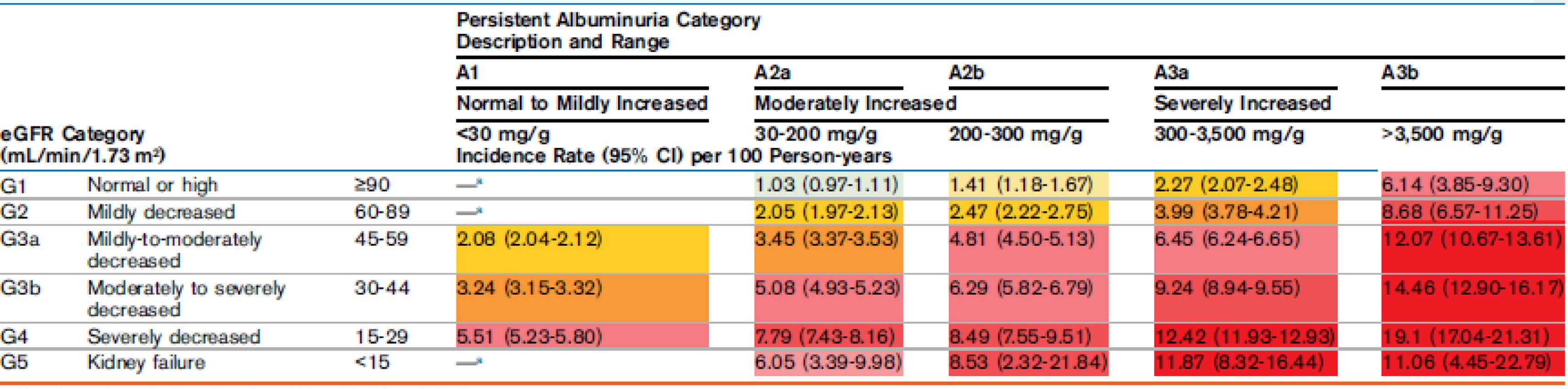
| Supplementary Table 2 Medical conditions and associated mechanisms contributing to hyperkalemia |
|
|---|---|
| Mechanism | Medical condition |
| Transcellular shift of K |
|
| Decreased/impaired K excretion |
|
| AKI, acute kidney injury; CKD, chronic kidney disease; CVD, cardiovascular disease. Data adapted from Larivee et al [25]. |
|
| Supplementary Table 3 Common medications associated with increased risk for hyperkalemia |
||
|---|---|---|
| Mechanisms | Class | Examples |
| Increased K load [29] | Potassium supplements | Slow K, K Dur |
| Decreased renal K excretion [11] | Angiotensin-converting enzyme inhibitor (ACEi) | Perindopril, ramipril, etc |
| Angiotensin receptor inhibitor (ARB) | Candesartan, irbesartan, etc | |
| Angiotensin receptor neprilysin inhibitor (ARNI) | Sacubitril/valsartan | |
| Direct renin inhibitor (DRI) | Aliskiren | |
| Aldosterone antagonist/mineralocorticoid receptor antagonist (MRA)/nsMRA | Spironolactone, eplerenone, finerenone | |
| Βeta-adrenergic receptor blocker | Metoprolol, atenolol, etc | |
| Heparin | Unfractionated heparin, low-molecular-weight heparin | |
| Nonsteroidal anti-inflammatory drug (NSAID) | Ibuprofen, celecoxib, etc | |
| Potassium-sparing diuretic | Amiloride, triamterene | |
| Calcineurin inhibitor (CNI) | Cyclosporine, tacrolimus | |
| Antimicrobials | Trimethoprim, pentamidine | |
| Impaired cellular K homeostasis [29] | Digitalis glycoside (impair cellular uptake of K) | Digoxin (overdose) |
| Nonselective beta-adrenergic receptor blocker (decrease cellular uptake of K) | Carvedilol, labetalol, etc | |
| Data adapted from Perazella [29] and Palmer [11]. | ||
Supplementary Table 4
Herbal supplements with potassium
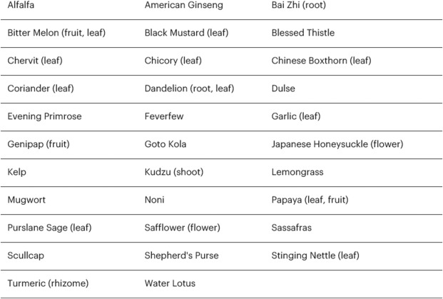
Supplementary Table 5
Potassium content of animal-origin foods, beverages, sweets, and fats
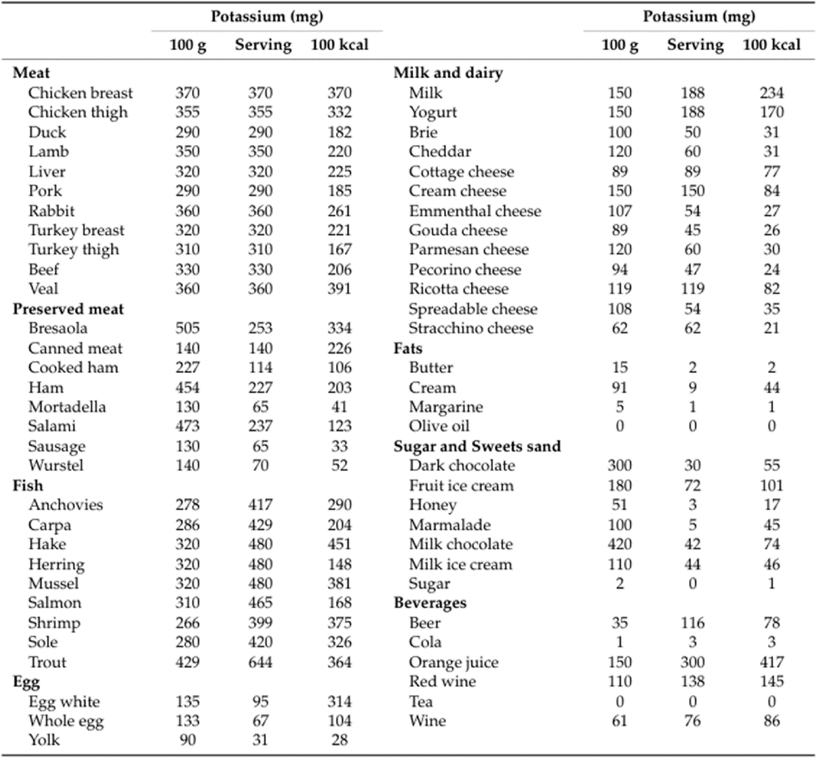
Note: 1 mmol of K = 39 mg [22].

| Supplementary Table 7 A comparison of potassium binders |
|||
|---|---|---|---|
| Potassium binders | |||
| Sodium polystyrene sulfonate (Kayexalate) | Sodium zirconium cyclosilicate (Lokelma) | Patiromer (Veltassa) | |
| Site of action | Colon | Small and large intestines | Colon |
| Exchange ion for K | Sodium | Sodium | Calcium |
| Onset of action | Variable, hours to days | 1 hour | 4–7 hours |
| Duration of effect | Variable; 6–24 hours | Not studied | 24 hours |
| Recommended dose | 15–30 g, 1–4 times per day orally, maximum 60 g/day | 10 g 1–3 times per day × 24–48 hours orally, maintenance therapy 5–10 g once per day | 8.4 g once per day orally, titrate up to maximum 25.2 g once per day |
| Adverse effects | GI symptoms (constipation, diarrhea, nausea, vomiting), serious GI effects (intestinal necrosis, bleeding, ischemic colitis, perforation)
|
GI symptoms (nausea, diarrhea, flatulence)
|
GI symptoms (constipation, diarrhea, nausea)
|
| Provincial coverage | https://prescribesmart.com | ||
| GI, gastrointestinal; K, potassium. Note: Data adapted from Weinstein et al [30,54]. |
|||
| Supplementary Table 8 Potassium measurement after medication changes for individuals with CKD and diabetes |
|
|---|---|
| Medication change | Suggested time frame for K recheck |
| Initiation or doubling of dose of: | |
|
1–2 weeks |
|
4 weeks |
| After K-binder therapy/ER visit for hyperkalemia | Within 1 week |
| Regular follow-up | 3–6 months |
| ARB, angiotensin receptor blocker; ACEi, angiotensin-converting enzyme inhibitor; CKD, chronic kidney disease; ER, emergency room; K, potassium; MRA, mineralocorticoid receptor antagonist; nsMRA, nonsteroidal mineralocorticoid receptor antagonist. | |
Supplementary Figure 1
Regulation of potassium homeostasis [47].
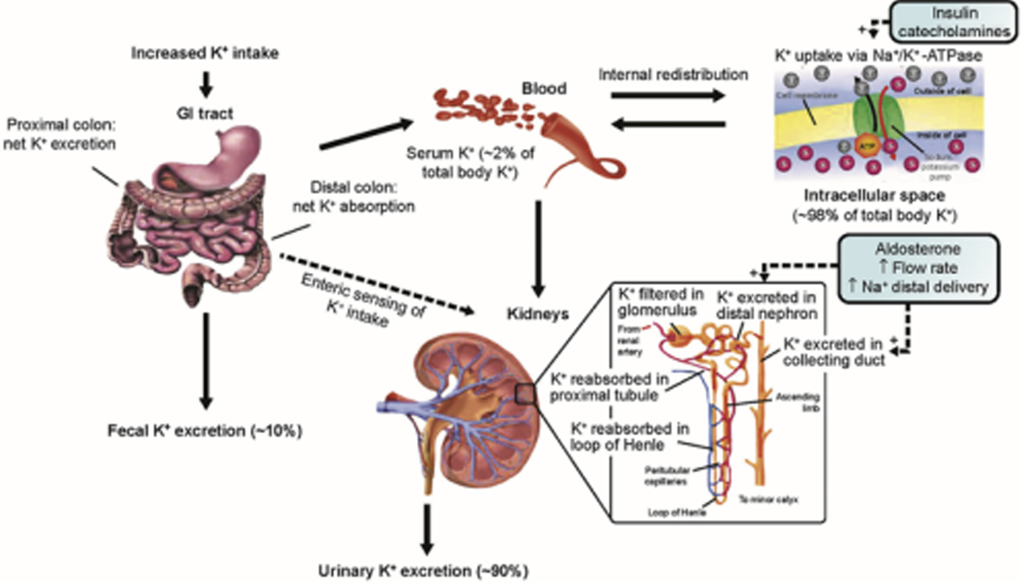
Supplementary Figure 2
Plants and herbal components with anti-arrhythmic activities and plants – cardiac medications interactions [32].

Supplementary Figure 3
Potassium absorption rates of different food groups [37].
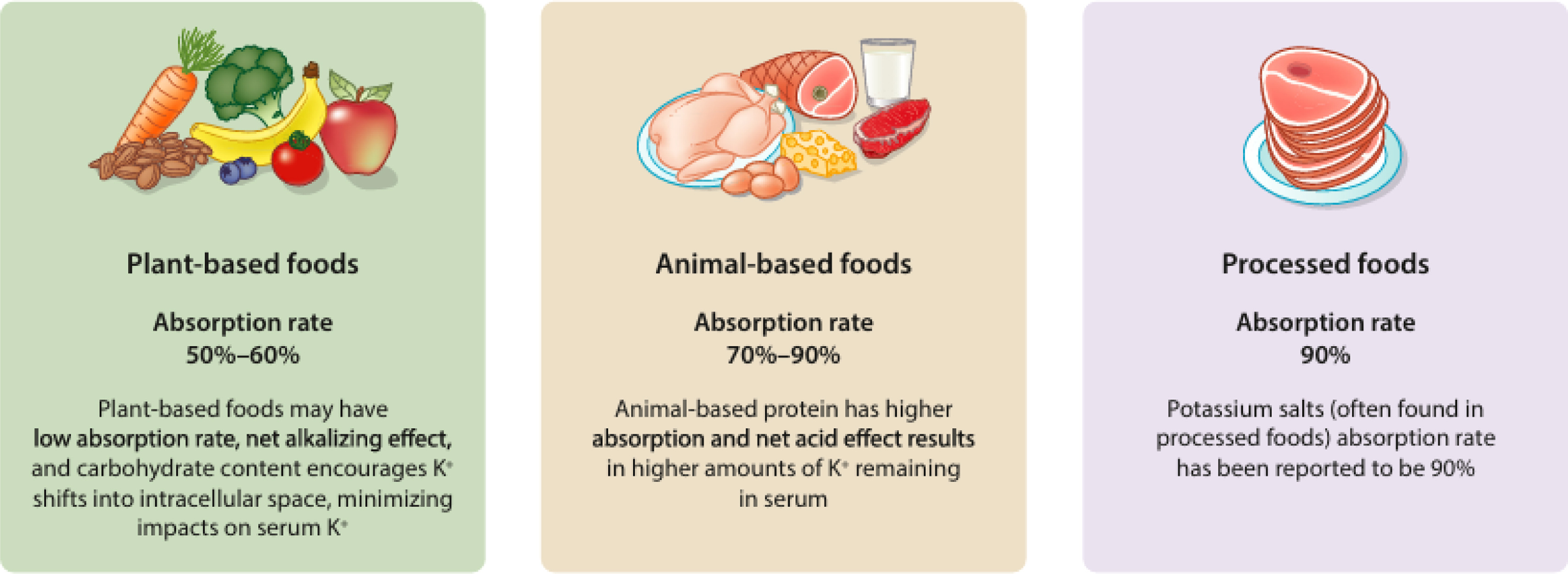
Supplementary Figure 4
Examples of processed/whole foods and processed with additives with high potassium contents. From BC Renal Dietitians Practice Group (http://www.bcrenal.ca/resource-gallery/documents/potassium_management_in_kidney_disease.pdf, accessed November 11, 2024).


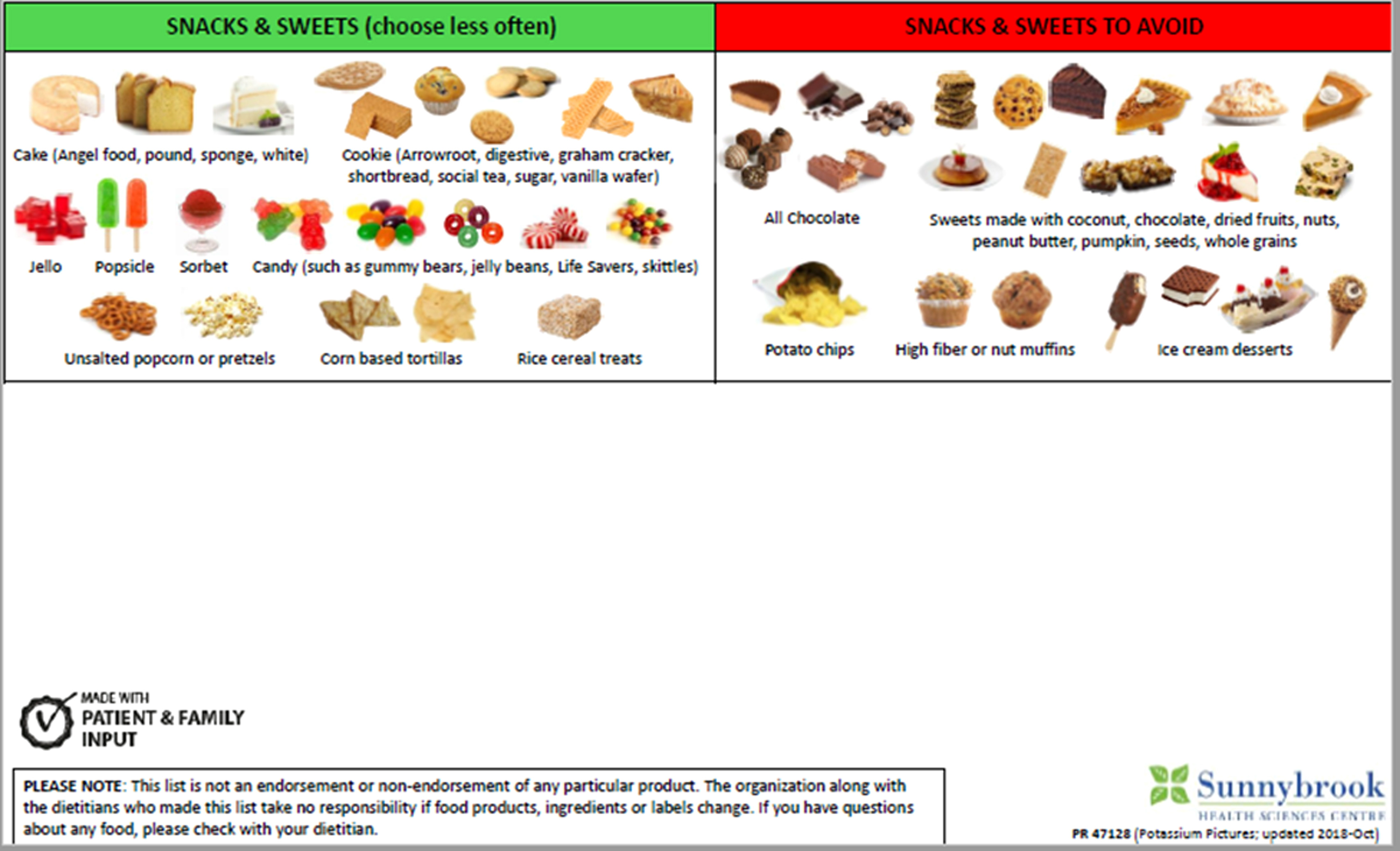
Supplementary Figure 6
Quick checklist of foods often associated with acute hyperkalemia and their typical potassium content.
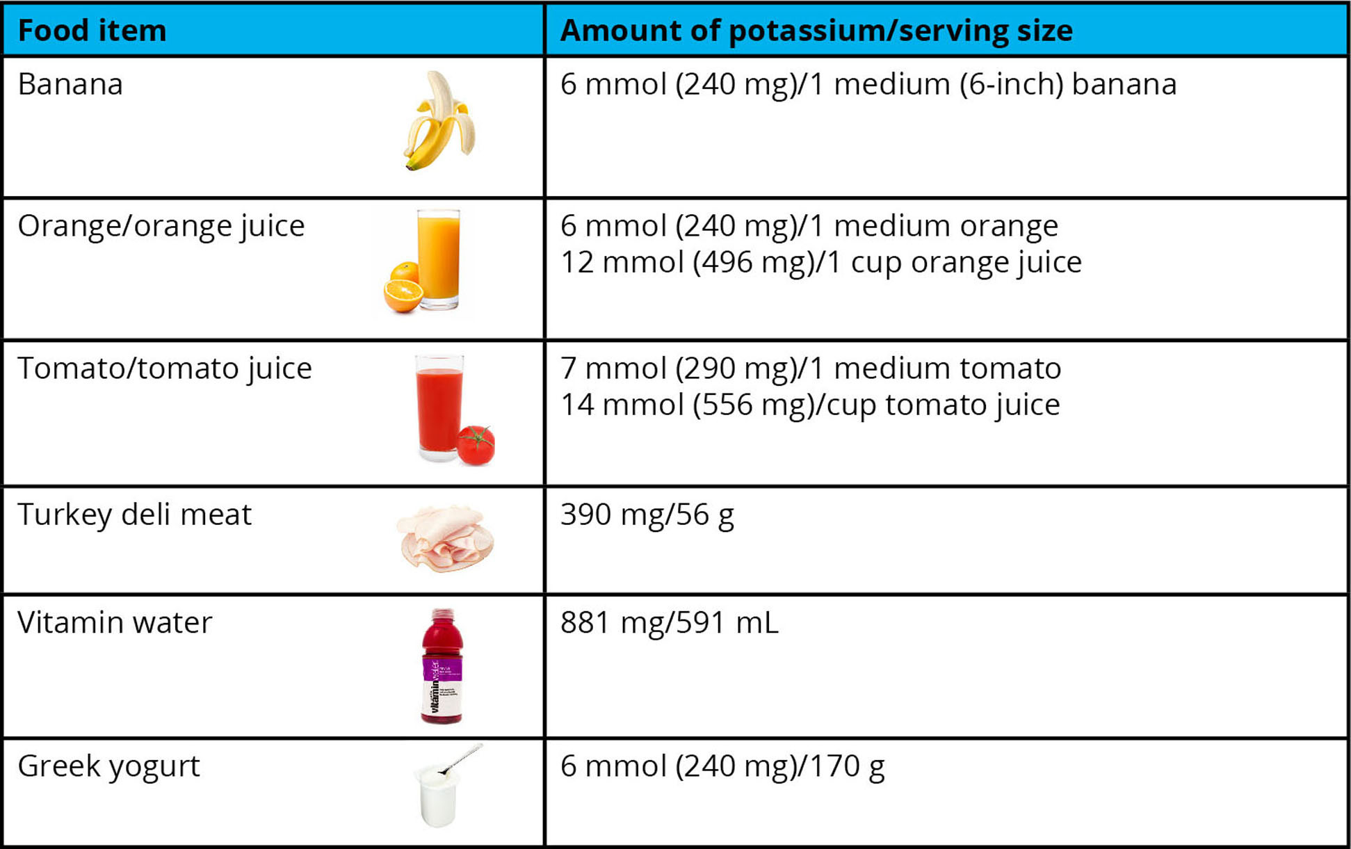
Supplementary Figure 7
Algorithm summarizing recommendations.

References
- Kovesdy CP. Management of hyperkalemia: An update for the internist. Am J Med 2015;128:1281e7.
- Hougen I, Leon SJ, Whitlock R, Rigatto C, Komenda P, Bohm C, et al. Hyper- kalemia and its association with mortality, cardiovascular events, hospitali- zations, and intensive care unit admissions in a population-based retrospective cohort. Kidney Int Rep 2021;6:1309e16.
- Jain N, Kotla S, Little BB, Weideman RA, Brilakis ES, Reilly RF, et al. Predictors of hyperkalemia and death in patients with cardiac and renal disease. Am J Cardiol 2012;109:1510e3.
- Adelborg K, Nicolaisen SK, Hasvold P, Palaka E, Pedersen L, Thomsen RW. Predictors for repeated hyperkalemia and potassium trajectories in high-risk patients—a population-based cohort study. PLoS One 2019;14:e0218739.
- Leon SJ, Whitlock R, Rigatto C, Komenda P, Bohm C, Sucha E, et al. Hyper- kalemia-related discontinuation of renin-angiotensin-aldosterone system inhibitors and clinical outcomes in CKD: A population-based cohort study. Am J Kidney Dis 2022;80:164e173.e1.
- Epstein M. Hyperkalemia constitutes a constraint for implementing renin- angiotensin-aldosterone inhibition: The widening gap between mandated treatment guidelines and the real-world clinical arena. Kidney International Supplements 2016;6(1):20e8.
- Howlett JG, Chan M, Ezekowitz JA, Harkness K, Heckman GA, Kouz S, et al. The Canadian Cardiovascular Society heart failure companion: Bridging guidelines to your practice. Can J Cardiol 2016;32:296e310.
- Rosano GMC, Tamargo J, Kjeldsen KP, Lainscak M, Agewall S, Anker SD, et al. Expert consensus document on the management of hyperkalaemia in patients with cardiovascular disease treated with renin angiotensin aldosterone sys- tem inhibitors: Coordinated by the Working Group on Cardiovascular Phar- macotherapy of the European Society of Cardiology. Eur Heart J Cardiovasc Pharmacother 2018;4:180e8.
- Alfonzo A. Clinical Practice Guidelines Treatment of Acute Hyperkalaemia in Adults. UK; 2020.
- Goia-Nishide K, Coregliano-Ring L, Rangel ÉB. Hyperkalemia in diabetes mellitus setting. Diseases 2022;10:20.
- Palmer BF, Clegg DJ. Diagnosis and treatment of hyperkalemia. Cleve Clin J Med 2017;84:934e42.
- Palmer BF, Clegg DJ. Physiology and pathophysiology of potassium homeo- stasis. Adv Physiol Educ 2016;40:480e90.
- Hunter RW, Bailey MA. Hyperkalemia: Pathophysiology, risk factors and consequences. Nephrol Dial Transplant 2019;34(Suppl. 3):iii2e11.
- Palmer BF, Clegg DJ. Physiology and pathophysiology of potassium homeo- stasis: Core curriculum 2019. Am J Kidney Dis 2019;74:682e95.
- Morales E, Cravedi P, Manrique J. Management of chronic hyperkalemia in patients with chronic kidney disease: An old problem with news options. Front Med 2021;8:653634.
- Sorensen MV, Matos JE, Praetorius HA, Leipziger J. Colonic potassium handling. Pflügers Arch Eur J Physiol 2010;459:645e56.
- Humphrey T, Davids MR, Chothia M-Y, Pecoits-Filho R, Pollock C, James G. How common is hyperkalaemia? A systematic review and meta-analysis of the prevalence and incidence of hyperkalaemia reported in observational studies. Clin Kidney J 2022;15:727e37.
- Borrelli S, De Nicola L, Minutolo R, Conte G, Chiodini P, Cupisti A, et al. Current management of hyperkalemia in non-dialysis CKD: Longitudinal study of patients receiving stable nephrology care. Nutrients 2021;13:942.
- Provenzano M, Minutolo R, Chiodini P, Bellizzi V, Nappi F, Russo D, et al. Competing-risk analysis of death and end stage kidney disease by hyper- kalaemia status in non-dialysis chronic kidney disease patients receiving stable nephrology care. J Clin Med 2018;7:499.
- Bakris G, Agiro A, Mu F, Cook EE, Greatsinger A, Sundar M, et al. Consequences of recurrent hyperkalemia on cardiovascular outcomes and mortality. JACC Adv 2024;3:101331.
- van Boemmel-Wegmann S, Bauer C, Schuchhardt J, Hartenstein A, James G, Pessina E, et al. Hyperkalemia incidence in patients with non-dialysis chronic kidney disease: A large retrospective cohort study from United States clinical care. Kidney Med 2024;6:100879.
- Cupisti A, Kovesdy CP, D’Alessandro C, Kalantar-Zadeh K. Dietary approach to recurrent or chronic hyperkalaemia in patients with decreased kidney func- tion. Nutrients 2018;10:261.
- Sumida K, Biruete A, Kistler BM, Khor B-H, Ebrahim Z, Giannini R, et al. New insights into dietary approaches to potassium management in chronic kidney disease. J Renal Nutr; 2023.
- Clase CM, Carrero J-J, Ellison DH, Grams ME, Hemmelgarn BR, Jardine MJ, et al. Potassium homeostasis and management of dyskalemia in kidney diseases: Conclusions from a Kidney Disease: Improving Global Outcomes (KDIGO) Controversies Conference. Kidney Int 2020;97:42e61.
- Larivée NL, Michaud JB, More KM, Wilson J-A, Tennankore KK. Hyperkalemia: prevalence, predictors and emerging treatments. Cardiol Ther 2023;12:35e63.
- Epstein M, Reaven NL, Funk SE, McGaughey KJ, Oestreicher N, Knispel J. Evaluation of the treatment gap between clinical guidelines and the utiliza- tion of renin-angiotensin-aldosterone system inhibitors. Am J Manag Care 2015;21(Suppl.):S212e20.
- Pantanowitz L. Drug-induced hyperkalemia. The American Journal of Medi- cine 2002;112(4):334.
- Hollander-Rodriguez JC, Calvert Jr JF. Hyperkalemia. Am Fam Physician 2006; 73:283e90.
- Perazella MA. Drug-induced hyperkalemia: Old culprits and new offenders. Am J Med 2000;109:307e14.
- Stevens PE, Ahmed SB, Carrero JJ, Foster B, Francis A, Hall RK, et al. KDIGO 2024 Clinical practice guideline for the evaluation and management of chronic kidney disease. Kidney Int 2024;105(Suppl.):S117e314.
- Kili´s-Pstrusin´ska K, Wiela-Hojen´ska A. Nephrotoxicity of herbal products in Europe—a review of an underestimated problem. Int J Mol Sci 2021;22: 4132.
- Beik A, Joukar S, Najafipour H. A review on plants and herbal components with antiarrhythmic activities and their interaction with current cardiac drugs. J Trad Complement Med 2020;10:275e87.
- St-Jules DE, Clegg DJ, Palmer BF, Carrero J-J. Can novel potassium binders liberate people with chronic kidney disease from the low-potassium diet?: A cautionary tale. Clin J Am Soc Nephrol 2022;17:467e72.
- MacLaughlin HL, McAuley E, Fry J, Pacheco E, Moran N, Morgan K, et al. Re- Thinking Hyperkalaemia Management in Chronic Kidney DiseasedBeyond Food Tables and Nutrition Myths: An Evidence-Based Practice Review. Nutrients 2024;16(1):3.
- Picard K, Mager D, Richard C. How food processing impacts hyperkalemia and hyperphosphatemia management in chronic kidney disease. Can J Diet Pract Res 2020;81:132e6.
- Picard K, Morris A. Potassium food additives and dietary management of serum potassium: proposed best-practice recommendations. J Renal Nutr; 2024.
- Picard K, Griffiths M, Mager DR, Richard C. Handouts for low-potassium diets disproportionately restrict fruits and vegetables. J Renal Nutr 2021;31:210e4.
- Greer RC, Marklund M, Anderson CAM, Cobb LK, Dalcin AT, Henry M, et al. Potassium-enriched salt substitutes as a means to lower blood pressure. Hypertension 2020;75:266e74.
- Rabi DM, McBrien KA, Sapir-Pichhadze R, Nakhla M, Ahmed SB, Dumanski SM, et al. Hypertension Canada’s 2020 comprehensive guidelines for the pre- vention, diagnosis, risk assessment, and treatment of hypertension in adults and children. Can J Cardiol 2020;36:596e624.
- Edwards A. Salt, salt substitutes, and seasoning alternatives. J Renal Nutr 2008;18:e23e5.
- Martínez-Pineda M, Vercet A, Yagüe-Ruiz C. Are food additives a really problematic hidden source of potassium for chronic kidney disease patients? Nutrients 2021;13:3569.
- Macdonald-Clarke CJ, Martin BR, McCabe LD, McCabe GP, Lachcik PJ, Wastney M, et al. Bioavailability of potassium from potatoes and potassium gluconate: A randomized dose response trial. Am J Clin Nutr 2016;104: 346e53.
- Chiu M, Garg AX, Moist L, Jain AK. A New Perspective to longstanding chal- lenges with outpatient hyperkalemia: A narrative review. Can J Kidney Health Dis 2023;10:20543581221149710.
- Liamis G, Liberopoulos E, Barkas F, Elisaf M. Diabetes mellitus and electrolyte disorders. World J Clin Cases 2014;2:488e96.
- Ferreira JP, Butler J, Rossignol P, Pitt B, Anker SD, Kosiborod M, et al. Abnor- malities of potassium in heart failure. J Am Coll Cardiol 2020;75:2836e50.
- Chang AR, Sang Y, Leddy J, Yahya T, Kirchner HL, Inker LA, et al. Antihyper- tensive medications and the prevalence of hyperkalemia in a large health system. Hypertension 2016;67:1181e8.
- Palmer BF, Carrero JJ, Clegg DJ, Colbert GB, Emmett M, Fishbane S, et al. Clinical management of hyperkalemia. Mayo Clin Proc 2021;1996:744e62.
- Lepage L, Dufour A-C, Doiron J, Handfield K, Desforges K, Bell R, et al. Ran- domized clinical trial of sodium polystyrene sulfonate for the treatment of mild hyperkalemia in CKD. Clin J Am Soc Nephrol 2015;10:2136e42.
- Harel Z, Harel S, Shah PS, Wald R, Perl J, Bell CM. Gastrointestinal adverse events with sodium polystyrene sulfonate (Kayexalate) use: A systematic review. Am J Med 2013;126(264):e9e24.
- Noel JA, Bota SE, Petrcich W, Garg AX, Carrero JJ, Harel Z, et al. Risk of hos- pitalization for serious adverse gastrointestinal events associated with sodium polystyrene sulfonate use in patients of advanced age. JAMA Intern Med 2019; 179:1025e33.
- Weir MR, Bakris GL, Bushinsky DA, Mayo MR, Garza D, Stasiv Y, et al. Patiromer in patients with kidney disease and hyperkalemia receiving RAAS inhibitors. N Engl J Med 2015;372:211e21.
- Kosiborod M, Rasmussen HS, Lavin P, Qunibi WY, Spinowitz B, Packham D, et al. Effect of sodium zirconium cyclosilicate on potassium lowering for 28 days among outpatients with hyperkalemia: The HARMONIZE randomized clinical trial. JAMA 2014;312:2223e33.
- Zannad F, Hsu B-G, Maeda Y, Shin SK, Vishneva EM, Rensfeldt M, et al. Efficacy and safety of sodium zirconium cyclosilicate for hyperkalaemia: The ran- domized, placebo-controlled HARMONIZE-Global study. ESC Heart Fail 2020; 7:55e65.
- Weinstein J, Girard L-P, Lepage S, McKelvie RS, Tennankore K. Prevention and management of hyperkalemia in patients treated with renineangiotensinealdosterone system inhibitors. CMAJ 2021;193:E1836e41.
- Sriperumbuduri S, McArthur E, Hundemer GL, Canney M, Tangri N, Leon SJ, et al. Initial and recurrent hyperkalemia events in patients with CKD in older adults: A population-based cohort study. Can J Kidney Health Dis 2021;8: 20543581211017408.
Diabetes Canada is the registered owner of all content on guidelines.diabetes.ca and ShopDC. For questions, please email info@diabetes.ca